Immunology

affinity maturation

Affinity maturation is a process of affinity-selected differentiation of activated B cells. Repeated exposures to the same antigen provokes greater antibody ligating affinity in the antibody secreted by successive generations of plasma cells.

The mechanisms by which affinity maturation is achieved are somatic hypermutation and clonal selection. Somatic hypermutation (SHM) is a diversity generating, regulated cellular mechanism through which antibodies are produced against an enormous variety of different potential antigens. The binding affinities of the variable regions of immunoglobulins are altered by AID-enzyme-promoted mutations during antigen-stimulated proliferation of B cells. These somatic hypermutations are transcribed and translated into thousands of slightly different immunoglobulins coded by the hypermutated V regions. The complementarity determining regions of these antibodies possess different affinities for the encountered antigen, and clonal selection will favor cells equipped with highest affinity antibodies because these B cells are favoured in terms of activation and co-operation with T cells.

Clonal selection is the phenomenon whereby a previously unencountered cognate antigen (epitope) can stimulate naïve B lymphocytes to proliferate and differentiate into clones of memory B cells and plasma cells that produce antibodies with the highest affinity for the antigen. Those B cells that have highest affinity BCR against the encountered antigen will be selected for proliferation, antibody production, and committment to an antigen-specific memory lineage.

Thus, SHM prepares a spectrum of antibodies with different affinities for the antigen, while clonal selection ensures that the immune system will react increasingly effectively (highest affinity) to an encountered antigen and will be ready for rapid response to subsequent encounters with the antigen.

Tables  Complement Receptors  Cytokines  Fc receptors  Immune Cytokines  Immunoglobulins  Interferons  Scavenger Receptors  Toll-like Receptors

tags

Labels: , , , , , , , ,

| 0 Guide-Glossary

anergy

Anergy (immunologic tolerance) refers to the failure to mount a full immune response against a target.

Anergy toward self-targets operates as one self-tolerance mechanism to control the autoreactive cells found in autoimmunity. Clonal deletion in which lymphocytes are killed if they recognize a self-antigen during their maturation in the thymus gland or bone marrow is a major mechanism for the prevention of autoimmunity. However, not all human self-antigens are expressed in the central lymphoid organs where the lymphocytes are developing. Thus, self-tolerance to an individual's own antigens must also depend on mechanisms such as clonal anergy. Theoretically, recognition of a self-antigen eliminates the proliferative capacity of autoreactive lymphocytes in the peripheral immune system. Another process, immunoregulation, utilizes regulatory T cells that weaken harmful or inappropriate lymphocyte responses.

In B cell anergy, self-reactive B cells persist in the periphery yet remain unresponsive to immunogen. Research findings indicate that continuous binding of antigen and subsequent receptor signaling are essential for the maintenance of anergy.[n]

T cell anergy is induced when TCR stimulation "freezes" T cell responses until they receive an adequate subsequent antigenic signal from an antigen-presenting cell. Such APC signals can rescue T cells from anergy, stimulating them to produce the lymphokines necessary for the growth of additional T cells.

During a productive immune response, CD4+ T cells respond to effective signals by producing interleukin 2 (IL-2) and by proliferating. Effective signals stimulate require both ligation of TCRs with cognate antigens presented by class II MHC molecules on the surface of APCs and activation of costimulatory receptors, such as CD28, which recognize ligands such as B7 proteins expressed on the surface of APCs.

When T cells receive stimulus only TCR signals in the absence of engagement of costimulatory receptors, they enter a state of anergic unresponsiveness characterized by an inability to produce IL-2 or to proliferate upon re-stimulation. Such anergic T cells show a profound block in Ras/MAPK pathway that prevents activation of the AP-1 family of transcription factors (Fos/Jun).

GRAIL (gene related to anergy in lymphocytes) is GRAIL is an E3 ubiquitin ligase that is necessary for the induction of CD4+ T cell anergy in vivo. It is upregulated in naturally occurring (thymically derived) CD4+ and CD25+ cells [a] and anergized T cells [1]. Both GRAIL and Foxp3 are genotypic marker for CD25+ Treg cells. T cell activation appears to be controlled by Foxp3 through transcriptional regulation of early growth response (Egr) genes Egr-2 and Egr-3, and E3 ubiquitin (Ub) ligase genes Cblb [?], Itch [?] and GRAIL, subsequently affecting degradation of two key signaling proteins, PLCgamma1 and PKC-theta. [a]

It is believed that GRAIL could induce anergy through ubiquitylation of membrane-associated targets required for T-cell activation. It has been demonstrated that two isoforms of otubain-1, in conjunction with the deubiquitylating enzyme USP8, produce opposing effects on the expression and function of GRAIL in the induction of anergy.[2] GRAIL is differentially expressed in naturally occurring and peripherally induced CD25+ Treg cells where the expression of GRAIL has been suggested is linked to their functional "regulatory" activity.

Tables  Complement Receptors  Cytokines  Fc receptors  Immune Cytokines  Immunoglobulins  Interferons  Scavenger Receptors  Toll-like Receptors

tags

Labels: , , , , , , , , , , , , , , , , ,

| 0 Guide-Glossary

APCs

APCs are antigen presenting cells, which display epitope proteins – exogenous antigen or fragmented antigen from phagocytosed cells – on their surfaces.

APC types : B cells : BCRs : CD1 family : CD1 proteins : CD4+ : class II MHC : dendritic cells : endocytosis : exocytosis : fragmented antigen peptides : histocompatibility molecules : γδ T cells : intact antigen : lipid antigen : macrophages : mycobacterial cell wall components : peptide antigen : phagocytic presenting cells : T cells and fragmented peptides, T cells and lipid antigens

Antigen presenting cells include:
phagocytic cells – dendritic cells, macrophages
B cells (B lymphocytes)
γδ T cells

Fragmented antigen – APCs engulf the antigen through endocytosis, then the endosome fuses with a lysosome where the antigen is digested into fragments such as short peptides. Following, exocytosis, a class II histocompatibility molecule holds the fragmented antigenic peptides at the surface of the cell, where they may be recognized by CD4+ T cells.

Intact antigen – dendritic cells can present intact antigen to B cells (not fragmented in lysosomes) by presenting the antigen on the cell surface. This antigen can bind to BCRs of the appropriate specificity, and can stimulate the B cells.

Presentation of peptide antigens for activation of naïve T cells does not reside solely in dendritic cells. A population of γδ T cells can efficiently present peptide antigens to αβT cells, and γδ T cells of the major tissue subset recognize self and foreign nonpeptide, lipid antigens presented by CD1 proteins. γδ T cells carry TCRs encoded by different gene segments than those of αβ T cells.

CD1 proteins are a family (CD1a-e) of cluster of differentiation glycoproteins related to the class I MHC molecules. CD1 are involved in the presentation of lipid and glycolipid antigens, particularly self, microbial, and mycobacterial cell wall components, to CD1-specific T cells.

The human CD1 family of transmembrane glycoproteins are encoded by five CD1 family genes organized in a cluster on chromosome 1. CD1 glycoproteins form heterodimers with beta-2-microglobulin. CD1 family members are considered to differ in cellular localization and specificity for particular lipid ligands. The CD1a protein (R4, T6, CD1, FCB6, HTA1) localizes to the plasma membrane and to recycling vesicles of the early endocytic system. Alternatively spliced transcript variants have been observed.[e]

ф activationalternative splicing ф antibodies ф antigen ф B cells cell membranes ф costimulation ф dendritic cellsendosomesexosome ф helper T cell ф killer T cellslysosome ф macrophages ф MHC ф pathogens ф pattern-recognition receptors ф phagocytephagocytosisreceptor-mediated endocytosis ф surface receptors ф γδ T cells ф T cells

Tables  Fc receptors  Immune Cytokines  Immunoglobulins  Cell Adhesion Molecules  Cell signaling  Receptor Tyrosine Kinases (RTKs)  Receptor Signal Transduction  Second Messengers 

tags

Labels: , , , , , , , , ,

| 0 Guide-Glossary

B cells

B cells are lymphocytes (WBCs) that participate in humoral immunity by producing antibodies in response to antigen stimulation.

activation : B-1 : B-2 : BCRs : CDRs : granzymes : helper T cells : life-span B cells : lymphopoiesis : memory B : naïve B cells : NK cells : NK receptors : NK cells attack viral infected cells : perforin : plasma B : stimulation : surface-immunoglobulins : surface receptors : VDJ recombination

Surface membrane-associated immunoglobulins (IgD and IgM) act as B cell receptors (BCRs), and the enormous variety of antigen recognition sites is attributable to VDJ recombination (alternative splicing) of peptide sequences encoded by V, D, and J genes. The variable region of immunoglobulins includes the recognition sites or complementarity determining regions (CDRs).
Lymphopoiesis, which takes place in the bone marrow of almost all mammals, produces small lymphocytes, large granular lymphocytes (NK) cells, B lymphocytes (precursors of plasma cells, T lymphocytes, and lymphoid dendritic cell. Recognition of self during lymphopoiesis permits anergy (suppression of self-attack).

Naïve B cells each have one of millions of distinct surface antigen-specific receptors, yet have not encountered their specific, cognate antigen. With a life-span of only a few days, many B cells die without ever encountering their cognate antigen. Naïve B cells are activated when the BCR binds to its cognate antigen. This antigen-Ig binding must be coupled with a signal from a helper T cell in order to activate the B cell.

Once activated, B lymphocytes:
● differentiate into one of the B cell types (directly or through intermediate, germinal center reactions)
● plasma cells produce antibodies against the antigenic stimulus, or memory cells are primed for subsequent activation by the antigen

Types of B cell:
B-1
B-2
Plasma B cells
Memory B cells

After newly formed B cells exit generative sites in fetal liver or adult bone marrow they undergo selection events that may involve interactions with self or with external antigens. Selective events can influence the phenotype and functional characteristics of B cells. B cell receptor-mediated events also influence lymphoid organs localization as marginal zone B cells in the spleen, as follicular (B-2 cells), as well as B-1 cells in the peritoneal and pleural cavities. [] fluorescence micrograph spleen, fm high power in which T cells form periarteriolar lymphocyte sheath (PALS) (red) and B-2 cell follicles (green) []

B-1 cells are the first B cells produced in the fetus, and in adults are located primarily in the peritoneal and pleural cavities. B1 cells are believed to operate in the innate response to infection by viruses and bacteria, and usually show preferential responses to T cell-independent antigens. The diversity of B-1 lymphocytes is attributed to their recombinatorial recombination, in which there is a preferential recombination between D-proximal VH gene segments. B-1 lymphocytes express (polyspecific) IgM in greater quantities than they express IgG, and the ability of B1 cells to respond to isotype switch commitment factors such as interleukin-4 may be secondary to their production of IgM. B-1 cells express CD5, which binds to CD72 to mediate B cell-B cell interactions.

B-2 cells are conventional B lymphocytes that are produced postnatally (unlike fetal B-1 cells) and are replaced from the bone marrow.

Plasma B lymphocytes are committed to production of copious amounts of monoclonal antibodies.

Memory B lymphocytes are long-lived, stimulated B lymphocytes that are primed for rapid response to a repeated exposure of the priming antigen. Memory B cells are generated in lymphoid tissue after B cell activation/proliferation and reside in the bone marrow, lymph nodes, and spleen. High affinity surface immunoglobulins enable their activation by lower levels of cognate antigen than are naïve B cells.

NK cells are differentiated from killer T cells. NK, natural killer cells constitute a corps of circulating lymphocytes that are constitutively specialized to attack cancerous cells and virus infected cells. Preprogramming for target recognition, coupled with the absense of need for backup by a clone of identical cells, renders NK cells capable of rapid (innate) response to pathogens. NK attack involves the exocytosis of cytoplasmic granules containing perforin and granzymes. Perforin forms pores in the plasma membrane of attacked cells through which serine-protease granzymes enter, cleaving caspase precursors and triggering apoptosis.

Individuals inherit multiple, polymorphic genes for NK receptors, so the assemblage of NK receptors differs between individuals. NK cells carry two forms of surface receptors:
● killer inhibitory receptors (KIRs) transmit an inhibitory signal when they encounter class I MHC molecules on a cell surface. (By contrast, T cells only recognize antigens that are presented by a MHC molecule.)
● activating receptors, which activate the NK cell upon binding to a target cell

Viral infection often causes suppression of MHC expresion, leading to a reduction of inhibition of NKs by its killer inhibitory receptors. This double negative renders the virus infected cell a target for killing by NK cells.

"About 85% of peripheral B cells are phenotypically mature and display first-order exponential kinetics defined by a half-life of 5-6 weeks, whilst the remainder are short-lived with a life span of several days."[s]

[] tem plasma cell [] micrograph macrophage surrounded by normal plasma cells [] micrograph macrophage & plasma cells []

activation : BCRs : CDRs : helper T cells : life-span B cells : lymphopoiesis : naïve B cells : surface-immunoglobulins : surface receptors : VDJ recombination

Tables  Fc receptors  Immune Cytokines  Immunoglobulins  Cell Adhesion Molecules  Cell signaling  Receptor Tyrosine Kinases (RTKs)  Receptor Signal Transduction  Second Messengers 

Top

tags

Labels: , , , , , ,

| 0 Guide-Glossary

CD

CD stands for cluster of differentiation, which indicates a defined subset of cellular surface receptors (epitopes) that identify cell type and stage of differentiation, and which are recognized by antibodies.

There are more than 250 identified clusters, each a different molecule, coating the surface of
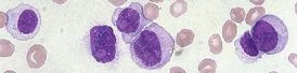
 B lymphocytes and T lymphocytes.

All T and B cells have about 105 = 100,000 molecules on their surface. B cells are coated with CD21, CD35, CD40, and CD45 together with non-CD molecules, while T cells express CD2, CD3, CD4, CD8, CD28, CD45R, and other non-CD molecules. Many CDs are expressed on both B and T cells, including CD5, CD6, CD23, CD27, CD28, CD84. Dendritic cells also express CD4 and CD8.

B lymphocyte CDs : B and T lymphocyte CDs : T lymphocyte CDs : TNFRs : CD1 : CD2 : CD3 : CD4 : CD5 : CD6 : CD8 : CD14 : CD23 : CD25 : CD27 : CD28 : CD30 : CD31 : CD36 : CD40 : CD45 : CD45 isoforms : CD55 : CD58 : CD72 : CD84 : complement CÆ : costimulatory : ITAMs : LFA : lymphocyte function associated antigen : glycoproteins : Ig superfamily : Macrophages/Monocytes : migration : pattern recognition receptors : platelets : scavenger receptors : SLAM (signaling lymphocyte activation molecule) : TNFR

T cell CDs:

CD2 family receptors are immunoglobulin (Ig) superfamily type I transmembrane, glycosylated proteins characterized by an N-terminal variable (V) domain that lacks disulfide bonds and a truncated Ig constant 2 (C2) domain that has two disulfide bonds in the extracellular region.

CD3 family receptors comprise three distinct chains – CD3γ, CD3δ and CD3ε in mammals – which are closely related proteins of the immunoglobulin superfamily, each containing a single extracellular immunoglobulin domain. The CD3 chains associate with TCRs and the ζ-chain to activate T lymphocytes. Together the TCR, ζ-chain, and CD3 molecules consitute the TCR complex.

The transmembrane region of the CD3 chains is negatively charged, enabling these chains to associate with the positively charged TCR chains (TCRα and TCRβ). The intracellular tails of the CD3 molecules contain a single conserved motif termed the immunoreceptor tyrosine-based activation motif (ITAM) that is essential for TCR signaling. Phosphorylation of CD3's ITAM enables the CD3 chain to bind Fyn, a membrane-associated protein tyrosine kinase important in the T cell's signaling cascade.

CD4 is notorious because of its importance in HIV/AIDs. It is an approximately 55 kDa type I membrane glycoprotein expressed predominantly on T cell precursors and a subset of mature T cells, providing the surface protein to which HIV attaches itself in order to invade the cell. CD4 is also found on the surface of monocytes, macrophages, Langerhans cells, astrocytes, keratinocytes and glial cells. The number of serum T4 cells is employed to measure the health of the immune system in people infected with HIV.[R&D]

CD8 (T8) is a protein embedded in the cell surface of 'suppressor' or regulatory T lymphocytes (Treg).

The CD31 adhesion molecule (PECAM-1) is expressed in large amounts at intercellular junctions of endothelial cells, subsets of T cells, platelets, and most other leukocytes including monocytes and neutrophils. CD31 is required for the trans-endothelial migration – extravasation – of leukocytes through intercellular junctions of vascular endothelial cells.

CD58 is also known as lymphocyte function-associated antigen (LFA-3). CD58 is a cell-bound immunoglobulin superfamily receptor with only one known ligand, which is CD2. CD58 is widely expressed on human hematopoietic and non-hematopoietic tissues, leukocytes, erythrocytes, endothelial and epithelial cells, and fibroblasts. The receptor-ligand pair, CD58 plus CD2, optimizes immune recognition and initiates T cell expansion and activation. Such contact activities can occur between helper T cells and antigen-presenting cells and between cytolytic effectors and target cells.

B and T cell CDs:

C5 is a 67 kDa surface glycoprotein of the scavenger receptor cysteine-rich (SRCR) superfamily that appears on thymocytes, mature T cells, and B cells. CD5 is important for the apoptosis of antigen-receptor induced B lymphocytes and for the maintenance of tolerance by anergic B cells. CD5 crosslinking induces extracellular mobilization of calcium ions, tyrosine phosphorylation of intracellular proteins, and production of diacylglycerol. Infection by EBV downregulates CD5 expression, while the glycoprotein is expressed in many T-cell leukemias and lymphomas.

CD6 is a member of the group B scavenger receptor cysteine-rich (SRCR) superfamily, which is expressed at low levels on immature thymocytes, at high levels on mature thymocytes, on the majority of peripheral blood T cells, a subset of B cells, and a subset of neuronal cells. Human and mouse CD6 proteins share 70% amino acid sequence identity over their full lengths.

CD23 is the receptor for the Fc portion of IgE.

CD27 Ligand/TNFSF7 is also known as CD70, and is a type II transmembrane glycoprotein belonging to the TNF superfamily (TNFSF). The expression of CD27 Ligand is induced by antigen-receptor activation in B cells. CD27/TNFRSF7 is a lymphocyte-specific member of the TNF receptor superfamily, which is expressed on a subset of human T cell precursors (thymocytes), on the majority of mature T cells, on natural killer (NK) cells, and subsets of B cells. CD27 ligation on NK cells induces proliferation and production of IFN-γ. CD27 binding (ligation) to T cells provides a co-stimulatory signal required for T cell proliferation, the promotion of effector T cell formation, and clonal expansion. The binding of CD27 to B cells inhibits the terminal differentiation of activated B cells into plasma cells and instead enhances commitment to memory B cell responses.

CD28 and CTLA-4, together with their ligands, B7-1 and B7-2, constitute one of the dominant B and T cell costimulatory pathways. CD28 and CTLA-4 are structurally homologous molecules of the immunoglobulin (Ig) gene superfamily. Mouse CD28 is expressed constitutively on almost all mouse T cells and on developing thymocytes.

CD45 is a protein tyrosine phosphatase (PTP) that regulates Src kinases required for T and B cell receptor signal transduction. CD45 dephosphorylates a negative regulatory residues on one or more of the protein tyrosine kinases that are involved in receptor-mediated second messenger formation. CD45 is located in all hematopoietic cells except erythrocytes and platelets, so it is also called the common leukocyte antigen.[]rotatable im[]

The CD45-regulated Src kinases are Lyn and Blk in B cells, and Lck and Fyn in T cells. ITAMs are immunocreceptor tyrosine bases motifs comprising two tyrosine residues separated by amino acids. RTK-phosphorylation of ITAMS enables them to bind to second family protein tyrosine kinases such as CD45, for which their SH2 domains have high binding affinity. In T cells, CD45 phosphorylates Csk, which is an inhibitory protein tyrosine kinase that controls tyrosine activity in lymphocytes. In B cells, calcium ions are transduced by the BCR, inducing CD45 expression. CD45RO, CD45RA, and CD45RB are isoforms of CD45.


CD84 is also known as Ly-9B, and is a member of the CD150/SLAM (signaling lymphocyte activation molecule) subfamily of the CD2 family (designated SLAMF5). CD84 is expressed on B cells, T cells, monocytes and platelets and acts as a self-ligand. Human and mouse CD84 share approximately 57% amino acid sequence identity.

B cell CDs:

CD40 is a type I transmembrane glycoprotein belonging to the TNF receptor superfamily. CD40 is expressed on B cells, follicular dendritic cells, dendritic cells, activated monocytes, macrophages, endothelial cells, vascular smooth muscle cells and several tumor cell lines. Human and mouse CD40s have 64% identity of amino acid sequence identity.


CD72 is a 39-43 kDa type II membrane glycoprotein of the C-type lectin family. CD72 is a pan-B cell marker that is expressed throughout the B lymphocytes differentiation (except plasma cells). CD72 is also present on follicular dendritic cells.

Monocytes/Macrophages

CD14 is a 55 kDa cell surface glycoprotein that is preferentially expressed on monocytes and macrophages. The amino acid sequence of human CD14 is approximately 65% identical to mouse CD14, and 82% identical to rat proteins.

Also: CD4, CD31 adhesion molecule (PECAM-1), CD40, CD84

Platelets:

CD36 is also known as scavenger receptor class B member 3 (SR-B3), GPIIIb, platelet membrane glycoprotein IV (GPIV), collagen receptor, thrombospondin receptor, and fatty acid translocase (FAT). CD36 is a broadly-expressed integral membrane glycoprotein with multiple physiological functions. As a member of the scavenger receptor family, CD36 is a multi-ligand pattern-recognition receptor that interacts with a large number of structurally dissimilar ligands. Upon ligand binding, CD36 transduces signals that mediate a wide range of pro-inflammatory cellular responses.

Complement CÆ activation family (RCA):

CD55, also known as decay-accelerating factor (DAF), is a 70 to 75 kDa member of the regulators of complement/CÆ activation (RCA) family of proteins. It is ubiquitously expressed on cells that are exposed to plasma complement proteins. Human CD55 is synthesized as a 381 amino acid precursor that comprises a 34 aa signal sequence, a 319 aa mature region and a 28 aa C-terminal prosegment.

Costimulatory:

CD28 and CTLA-4, together with their ligands, B7-1 and B7-2.

Pattern-recognition receptors :

CD36

Glycoproteins:

CD4, CD5, CD14, CD27, CD30, CD36, CD40, CD72

Immunoglobulin superfamily:

CD2, CD58, CD28 and CTLA-4,
CD23 is the receptor for the Fc portion of IgE.

LFA (lymphocyte function associated antigen):

CD58

migration:

CD31 adhesion molecule (PECAM-1)

Scavenger receptor (SRCR) family:

CD5, CD6, CD36

SLAM (signaling lymphocyte activation molecule) subfamily:

CD84 (Ly-9B), CD2 family (SLAMF5)


TNFRs :

CD30/TNFRSF8 is a type I transmembrane glycoprotein belonging to the TNF receptor superfamily, where the the ligand for CD30 is CD30L (CD153, TNFSF8), which is a member of the TNF superfamily. CD30 binding by CD30L mediates pleiotropic effects, including cellular proliferation, activation, differentiation, and apoptosis.

Other TNFRs are CD40 and CD27.

Miscellaneous CDs
CD9 is a member of the tetraspanin transmembrane receptor family. CD9 contains four putative transmembrane domains and two extracellular loops, and is thought to be involved in egg-sperm fusion. Several reports indicate that CD9 associates with integrins and affects cell behavior on fibronectin surfaces (ref).[s]

B lymphocyte CDs : B and T lymphocyte CDs : T lymphocyte CDs : TNFRs : CD2 : CD3 : CD4 : CD5 : CD6 : CD8 : CD14 : CD23 : CD27 : CD28 : CD30 : CD31 : CD36 : CD40 : CD45 : CD45 isoforms : CD55 : CD58 : CD72 : CD84 : complement CÆ : costimulatory : ITAMs : LFA : lymphocyte function associated antigen : glycoproteins : Ig superfamily : Macrophages/Monocytes : migration : pattern recognition receptors : platelets : scavenger receptors : SLAM (signaling lymphocyte activation molecule) : TNFR

Top

[C][CD5][CD72] Tables  Fc receptors  Immune Cytokines  Immunoglobulins


tags

Labels: , , , , , , , , , , , , , , , ,

| 0 Guide-Glossary

hematopoiesis

Hematopoiesis is the production of blood cells, a developmental process located in the (red) bone marrow, though some cells mature elsewhere. For example, T lymphocytes are so named because they mature in the thymus, and antigenic stimulation of B lymphocytes to become plasma cells typically takes place in the periphery.

B lymphocyte development : common lymphoid progenitor : common myeloid progenitor : E2A : EBF : early B lineage : erythropoiesis : granulopoiesis : hematopoietic growth factors : lymphopoiesis : monocytopoiesis : Pax-5 : pluripotential stem cell : precursors : progenitors : regulatory transcription factors : stages : stem cells : thrombopoiesis : transcriptional regulatory proteins

The process of haematopoiesis occurs in several stages, and is controlled by at least 11 hematopoietic growth factors (including the colony-stimulating factors, IL-2 through IL-7, G-CSF, GM-CSF, and M-CSF). The first stage involves the differentiation of a pluripotential stem cell into a committed progenitor, which is followed by maturation of committed progenitors in distinct pathways, in which precursors are partially developed, 'adolescent' cells en route to maturity.

stem → progenitor → precursor → adult → mature

[] labeled photomicrograph of bone marrow, diagram of adult stem cell plasticity, diagram of stem cell versus progenitor cell

Stem cell stage:
pluripotential hematopoietic stem cell
--------------------

The common myeloid progenitor can generate:

● proerythroblasts (pronormoblasts) → erythropoiesis

● myeloblasts → granulopoiesis

● monoblasts → monocytopoiesis

● megakaryoblasts → thrombopoiesis

The common lympoid progenitor can generate:

● lymphoblasts → lymphopoiesis

۝

Committed progenitor stage to mature cell : granulopoiesis

common myeloid progenitor

myeloblast

B/E/N promyelocyte

------

B/E/N myelocyte

------

B/E/N metamyelocyte

------

B/E/N band

------

basophil, eosinophil, neutrophil

-----------------------

mast cell -----------------------

۝

Committed progenitor stage to mature cell : lymphopoiesis

common lymphoid progenitor

lymphoblast

----------------------------------------↓ rearrangements H: D-J → H: V-DJ

prolymphocyte

-----------------↓ rearrangements L: V-J --------------------------

small lymphocyte------or----- natural killer cell (large granular lymphocyte)

------------↓ IgM→IgD ---------------------------------------------

------B lymphocyte--or-- T lymphocyte

-----------------------------------------------------------------------

--------plasma cell------------------------------------------lymphoid dendritic cell

Development of mature B lymphocytes from multipotent progenitors requires the coordinated activities of a number of transcriptional regulatory proteins, including EBF, Pax-5, and E2A.

During B cell-development from the precursor stage, differentiation involves rearrangement of the heavy chain gene segments. The functional integrity of the rearranged gene is tested: Precursor-B cells express two single domain Ig-like proteins of invariant sequence that substitute for the light chain. Formation of a complex comprising the μ (mu) heavy chain with the surrogate light chains instructs the cell to discontinue rearrangement of the heavy chain locus and to commence rearrangement of the k (kappa) locus. If successful light chain rearrangement is achieved such that the light and heavy chains form a complete antibody, then this complex instructs the cell to discontinue rearrangement of light chains, ensuring that only a single specificity is produced (allelic exclusion). Џ B cell maturation - animation Џ

Those developing B cell clones that fail to generate a productive rearrangement at both one of their heavy chain alleles and a light chain locus will undergo apoptosis. Immune tolerance mechanisms also exist to ensure the death of any newly produced B cells that express an antibody that reacts strongly with self proteins on the surface of host cells.

E2A proteins function in early B lineage development to regulate B lineage-specific gene expression as well as B cell survival. E2A-encoded proteins are involved in the differentiation of a number of cell types, and they are especially important in lymphocyte development.

The E2A gene encodes E47 and E12, which are basic-helix-loop-helix (bHLH) transcription factors that bind DNA either as homodimers or as heterodimers with other bHLH proteins. Such bHLH DNA binding activity in the B-lineage comprises E47 homodimers. Development of thymocytes mainly involves heterodimers of E47 and a related bHLH protein, HEB. Thymocytic E2A protein expression is required to initiate T-cell differentiation. During the development of thymoctyes, E-proteins and their antagonists, Id2 and Id3, regulate T-lineage specific gene expression and TCR rearrangement. E2A and Id proteins block thymocytic maturation in the absence of pre-TCR expression, and pre-TCR signaling acts to promote development in part by inhibiting E2A activity. [l]

۝

Committed progenitor stage to mature cell : monocytopoiesis

common myeloid progenitor

monoblast

promonocyte

monocyte

----

macrophage or myeloid dendritic cell


۝

Committed progenitor stage to mature cell : erythropoiesis

common myeloid progenitor

proerythroblast

basophilic erythroblast

polychromatic erythroblast

polychromatic erythrocyte (reticulocyte)

erythrocyte (RBC)

۝

Committed progenitor stage to mature cell : thrombopoiesis

megakaryoblast
promegakaryocyte
megakaryocyte
thrombocytes (platelets)

Top


tags

Labels: , , , ,

| 0 Guide-Glossary

humoral immunity

The humoral immune response employs antibodies secreted by B lymphocytes, and is a component of the adaptive immune response. In addition to antibody production, the immune system secretes chemical mediators.

Elements of the humoral response include:
● secretion of antibodies
___● neutralization of pathogens and toxins
___● activation of classical complement pathway
___opsonin production
● stimulation of cellular responses
___● formation of germinal centers
___activation of helper T cells (Th2)
___● production of memory B lymphocytes
___opsonin-directed phagocytosis and elimination of pathogens
● chemi-genetic responses
___isotype switching between immunoglobulin types
___affinity maturation (combining somatic hypermutation and affinity based clonal selection)
___● production of immune cytokines
___C9 complement membrane attack complex
___● production of acute phase-inflammatory response mediators

Tables  Complement Receptors  Cytokines  Fc receptors  Immune Cytokines  Immunoglobulins  Interferons  Scavenger Receptors  Toll-like Receptors

tags

Labels: , , , , ,

| 0 Guide-Glossary

lymphoid system

Components of the lymphoid system are:
● immune cells – B cell lymphocytes and plasma cells, dendritic cells, granulocytes, macrophages, monocytes of mononuclear phagocyte system (MPS), T cell lymphocytes
● lymph ducts and vessels and lymph nodes (right)
[] histopathology []
● lymphoid organs including reticuloendothelial system – bone marrow, (lymph nodes), mucosa-associated lymphoid tissue, Peyer's patches, spleen, thymus, tonsils, vermiform appendix

The reticuloendothelial system or mononuclear phagocytic system comprises a range of cells that are capable of phagocytosis, including macrophages and monocytes. Phagocytosis is an innate immune process, and is not an adaptive immune process. The phagocytic cells either circulate in the blood or are attached to various connective tissues such as pulmonary alveoli, liver sinusoids, skin, spleen, and joints.

The RES functions to provide phagocytic cells for both the inflammatory response and immune responses (primary RES) and to remove pathogens and senescent cells from circulation (secondary RES)

The reticuloendothelial system (RES) includes:
● primary (central) lymphoid production organs – bone marrow, thymus
● secondary (peripheral) lymphoid function organs – circulating monocytes, histiocytes located in many tissues, Kupffer cells of the liver, "Littoral cells" of the spleen, mucosa-associated lymphoid tissue (MALT), which is subdivided into bronchus-associated lymphoid tissue (BALT) and gut-associated lymphoid tissue (GALT), Peyer's patches.

Tables  Fc receptors  Immune Cytokines  Immunoglobulins

o-o index of tissue micrographs [] germinal centers [] fluorescence microscopy dendritic cell uptake of dying cells within spleen [] micrograph red pulp of spleen [] micrograph splenic red pulp [] micrograph spleen cells (mouse) DAPK2 stained [] micrograph white pulp, splenic nodule [] micrograph white pulp infiltrate with Langhans giant cell [] histopathology spleen Gaucher's disease [] micrograph gallery cell surface antigens [] Virtual Histology Main []


Top

Labels: , , , , , , , ,

| 0 Guide-Glossary

plasma cells

Plasma cells are B lymphocytes that have been activated to differentiate and mature by CD4+ helper T lymphocytes. Activated B cells become either memory B cells or plasma cells, which secrete copious amounts of monoclonal antibodies against the original antigen that triggered the antigen presenting cells.

Affinity maturation is a process of affinity-selected differentiation and maturation of activated B cells. Repeated exposures to the same antigen provokes greater antibody ligating affinity in the antibody secreted by successive generations of plasma cells. Isotype switching, in response to signaling by specific cytokines, provides for a switch of production from IgM to other Ig isotypes following first exposure to an antigen. T cell-produced immune cytokines, such as interleukin-4 (IL-4), interferon-γ (IFN-γ), and TGF-β effect isotype switching.

Plasma cells are predominantly located in the bone marrow (0.2% to 2.8% of leukocytes) and are rarely found in the peripheral blood.(description of morphology)

Plasma cells are seen in abnormal numbers in multiple myeloma, plasma cell leukemia, Waldenström's macroglobulinemia, and MGUS (monoclonal gammopathy of uncertain significance).

[] tem plasma cell [] micrograph macrophage surrounded by normal plasma cells [] micrograph macrophage & plasma cells []

Tables  Fc receptors  Immune Cytokines  Immunoglobulins

Labels: , , , , , , , ,

| 0 Guide-Glossary

. . . since 10/06/06
Google