Immunology

antibodies

Antibodies are glycoproteins of the immunoglobulin superfamily, and are adhesion-signaling molecules that recognize (bind to) specific antigens. Antibodies are synthesized by B cell-derived plasma cells.

▼: adhesion molecules : antigen binding site : C : CH1-4 : cellular adhesion molecules : complement fixation : complementarity determining regions : constant domains : domains : evolution of immunoglobulins : Fab : Fc : heavy chain : hinge region : Ig supergene family : isotypes : kinase activation : light chain : location of Ig classes : membrane-bound Igs : multimeric structures : tissue location : V : VDJ recombination : VH, VL : variable domains :▼
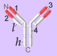
Immunoglobulins (left - click to enlarge) comprise two heavy (h) and two light-chain (l) protein subunits, each of which folds into domains (4 on heavy, 2 on light). These adhesion sites or domains contain one or more folds of 60 to 100 amino acids.

Depending upon the character of the heavy chain, immunoglobulins are divided into five classes – IgG, IgD, IgE, IgA, IgM – that are expressed in different tissues. The classes are further subdivided into isotypes, which have different properties in terms of complement fixation and binding to immunoglobulin (Ig) receptors.

Members of the immunoglobulin supergene family are found as:
● membrane-bound surface receptors of immune-system cells,
cellular adhesion molecules, or
● soluble antibodies (γ-globulins) synthesized by activated B cells.

Membrane-bound Igs have a transmembrane segment and a cytoplasmic C-terminal tail. The 2 β- chains are stabilized into sandwiched β sheets that are adherent by virtue of hydrophobic interactions between disulphide bonds. Igs assume a Y-shaped structure "topped" at the extracellular N-terminals by variable domains (red), with a variable domain at the tip of the heavy chain (1) and the light chain (2), between which lies an antigen binding site (3). The variable regions are coded by pluripotential DNA sequences that can generate thousands of polypeptide sequences capable of adhering to millions of different ligands. Binding is homophilic or heterophilic, including binding to different Igs and to integrins. Both light and heavy chains contain constant domains (white, 4).
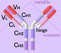
Right - click to enlarge - the heavy chains of IgA, IgD and IgG each have four domains, where those at the N-terminal are variable (VH) and the other three are constant (CH1-3). IgE and IgM have one variable and four constant domains (CH1-4) on the heavy chain. The variable domains are termed Fab, while the constant domains are termed Fc.

The light chains have two domains, one variable domain (VL) at the N-terminal, and one constant (CL) domain.

The antigen binding site lies between VH and VL (shaded lavendar). Most variability is found in three superficial-loop forming regions in the VH and VL domains, which are the complementarity determining regions or CDRs. CDR3 binds antigens and CDR1-2 bind MHCs. CDR3 shows more variation that do either CDR1 or 2.

The domains have related amino acid sequences that possess a common secondary and tertiary structure. This conserved structure is found frequently in proteins involved in cell-cell interactions and is particularly important in immunology. The constant (Fc) regions have complement fixing and Ig receptor binding activity. The hinge region, in IgG, IgA and IgD, is an important sequence of 10-60 amino acids between CH1 and CH2 that confers flexibility on the molecule.

animations Џ B cell selection Џ ELISA test +ve, -ve Џ IgG rotating x- y- axes Џ Rotating mouse IgG2a Molecule (y-axis) Џ somatic recombination of Ig gene Џ spinning IgG1 Kol Џ unfolding (small) IgG . unfolding (large) IgG .

Immunoglobulins attain their enormous variability by splicing components (VDJ recombination) coded in widely scattered sequences of DNA that are located in two different chromosomes. Antigen binding takes place at the heavy chain, which displays enormous variation by virtue of combining 1 of 400 possible variable gene segments with 1 out of 15 diversity segments and 1 out of 4 joining segments. This alternative splicing generates 24,000 possible combinations for the DNA encoding the heavy chain alone. The variable coding segments are assembled together with those for the constant-C segments of the heavy-chain molecule.

Tissue location:
IgA – mucus – gut, respiratory tract
IgD – antigen receptor on B cells
IgE – mast cells – releases histamines in response to allergens
IgG – primary immunity against invading pathogens
IgM – early B cell-mediated response to invading pathogens

Some antibody classes form multimeric structures – pentamers (IgM) and dimers or trimers (IgA). These two isotypes also associate with a small protein called the joining (J) chain required for stabilisation of the complexes.

The immunoglobulin superfamily is evolutionarily ancient, is widely expressed, and is constitutive or long-term up-regulated. Immunoglobulin antibodies are released by activated B cells of the immune system, on which they also act as surface marker proteins. Adherence of immunoglobilins to foreign substances or to cellular invaders may be sufficient to disarm the invader, or the attached antibodies function as attack signal to macrophages and natural killer cells. Adhesion molecules of the immunoglobulin supergene family, activate specific kinases through phosphorylation, resulting in activation of transcription factors, increased cytokine production, increased cell membrane protein expression, production of reactive oxygen species, and cell proliferation.

▲: adhesion molecules ~ adhesion molecules ф antigen : antigen binding site ф APCs ф B cells : C : CH1-4 : cellular adhesion molecules : complement fixation ф complement system : complementarity determining regions : constant domains : domains : evolution of immunoglobulins : Fab : Fc : heavy chain : hinge region ф humoral immunity : Ig supergene family ~ immunoglobulins : isotypes : kinase activationkinases : light chain : location of Ig classes : membrane-bound Igs : multimeric structures ф receptors ф signaling ф surface receptors ф T cells : tissue location ~ tyrosine kinases : V : VDJ recombination ф VDJ recombination : VH, VL : variable domains :▲

Tables  Fc receptors  Immune Cytokines  Immunoglobulins  Cell Adhesion Molecules  Cell signaling  Receptor Tyrosine Kinases (RTKs)  Receptor Signal Transduction  Second Messengers 

Top

tags

Labels: , , , , , , , , , ,

| 0 Guide-Glossary

B cells

B cells are lymphocytes (WBCs) that participate in humoral immunity by producing antibodies in response to antigen stimulation.

activation : B-1 : B-2 : BCRs : CDRs : granzymes : helper T cells : life-span B cells : lymphopoiesis : memory B : naïve B cells : NK cells : NK receptors : NK cells attack viral infected cells : perforin : plasma B : stimulation : surface-immunoglobulins : surface receptors : VDJ recombination

Surface membrane-associated immunoglobulins (IgD and IgM) act as B cell receptors (BCRs), and the enormous variety of antigen recognition sites is attributable to VDJ recombination (alternative splicing) of peptide sequences encoded by V, D, and J genes. The variable region of immunoglobulins includes the recognition sites or complementarity determining regions (CDRs).
Lymphopoiesis, which takes place in the bone marrow of almost all mammals, produces small lymphocytes, large granular lymphocytes (NK) cells, B lymphocytes (precursors of plasma cells, T lymphocytes, and lymphoid dendritic cell. Recognition of self during lymphopoiesis permits anergy (suppression of self-attack).

Naïve B cells each have one of millions of distinct surface antigen-specific receptors, yet have not encountered their specific, cognate antigen. With a life-span of only a few days, many B cells die without ever encountering their cognate antigen. Naïve B cells are activated when the BCR binds to its cognate antigen. This antigen-Ig binding must be coupled with a signal from a helper T cell in order to activate the B cell.

Once activated, B lymphocytes:
● differentiate into one of the B cell types (directly or through intermediate, germinal center reactions)
● plasma cells produce antibodies against the antigenic stimulus, or memory cells are primed for subsequent activation by the antigen

Types of B cell:
B-1
B-2
Plasma B cells
Memory B cells

After newly formed B cells exit generative sites in fetal liver or adult bone marrow they undergo selection events that may involve interactions with self or with external antigens. Selective events can influence the phenotype and functional characteristics of B cells. B cell receptor-mediated events also influence lymphoid organs localization as marginal zone B cells in the spleen, as follicular (B-2 cells), as well as B-1 cells in the peritoneal and pleural cavities. [] fluorescence micrograph spleen, fm high power in which T cells form periarteriolar lymphocyte sheath (PALS) (red) and B-2 cell follicles (green) []

B-1 cells are the first B cells produced in the fetus, and in adults are located primarily in the peritoneal and pleural cavities. B1 cells are believed to operate in the innate response to infection by viruses and bacteria, and usually show preferential responses to T cell-independent antigens. The diversity of B-1 lymphocytes is attributed to their recombinatorial recombination, in which there is a preferential recombination between D-proximal VH gene segments. B-1 lymphocytes express (polyspecific) IgM in greater quantities than they express IgG, and the ability of B1 cells to respond to isotype switch commitment factors such as interleukin-4 may be secondary to their production of IgM. B-1 cells express CD5, which binds to CD72 to mediate B cell-B cell interactions.

B-2 cells are conventional B lymphocytes that are produced postnatally (unlike fetal B-1 cells) and are replaced from the bone marrow.

Plasma B lymphocytes are committed to production of copious amounts of monoclonal antibodies.

Memory B lymphocytes are long-lived, stimulated B lymphocytes that are primed for rapid response to a repeated exposure of the priming antigen. Memory B cells are generated in lymphoid tissue after B cell activation/proliferation and reside in the bone marrow, lymph nodes, and spleen. High affinity surface immunoglobulins enable their activation by lower levels of cognate antigen than are naïve B cells.

NK cells are differentiated from killer T cells. NK, natural killer cells constitute a corps of circulating lymphocytes that are constitutively specialized to attack cancerous cells and virus infected cells. Preprogramming for target recognition, coupled with the absense of need for backup by a clone of identical cells, renders NK cells capable of rapid (innate) response to pathogens. NK attack involves the exocytosis of cytoplasmic granules containing perforin and granzymes. Perforin forms pores in the plasma membrane of attacked cells through which serine-protease granzymes enter, cleaving caspase precursors and triggering apoptosis.

Individuals inherit multiple, polymorphic genes for NK receptors, so the assemblage of NK receptors differs between individuals. NK cells carry two forms of surface receptors:
● killer inhibitory receptors (KIRs) transmit an inhibitory signal when they encounter class I MHC molecules on a cell surface. (By contrast, T cells only recognize antigens that are presented by a MHC molecule.)
● activating receptors, which activate the NK cell upon binding to a target cell

Viral infection often causes suppression of MHC expresion, leading to a reduction of inhibition of NKs by its killer inhibitory receptors. This double negative renders the virus infected cell a target for killing by NK cells.

"About 85% of peripheral B cells are phenotypically mature and display first-order exponential kinetics defined by a half-life of 5-6 weeks, whilst the remainder are short-lived with a life span of several days."[s]

[] tem plasma cell [] micrograph macrophage surrounded by normal plasma cells [] micrograph macrophage & plasma cells []

activation : BCRs : CDRs : helper T cells : life-span B cells : lymphopoiesis : naïve B cells : surface-immunoglobulins : surface receptors : VDJ recombination

Tables  Fc receptors  Immune Cytokines  Immunoglobulins  Cell Adhesion Molecules  Cell signaling  Receptor Tyrosine Kinases (RTKs)  Receptor Signal Transduction  Second Messengers 

Top

tags

Labels: , , , , , ,

| 0 Guide-Glossary

isotype switching

Isotype switching is a characteristic feature of the humoral immune response, in which a switch from IgM to other Ig isotypes follows first exposure to an antigen. Affinity maturation ensures that repeated exposures to the same antigen will provoke greater antibody ligating affinity of the antibody secreted by successive generations of plasma cells.

Isotype switching is regulated by T cell-produced immune cytokines, such as interleukin-4 (IL-4), interferon-γ (IFN-γ), and TGF-β, which direct B cells to switch to specific Ig classes.

Tables  Fc receptors  Immune Cytokines  Immunoglobulins

Labels: , , , , , ,

| 0 Guide-Glossary

MHC

The major histocompatibility complex (MHC) encodes molecules displayed on cell surfaces, where they participate in lymphocyte recognition and antigen presentation.

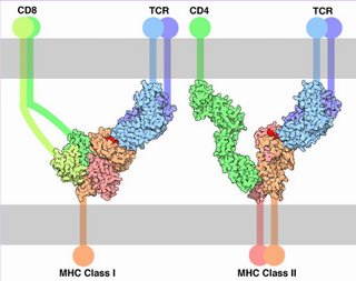
Left - click to enlarge - the presented antigen of the MHC-peptide complex is indicated by red. (Image source Molecule of Month: Illustration by David S. Goodsell of The Scripps Research Institute. Original work of the US Federal Government - public domain.)

The TCR and MHC-peptide complex make primary contact, then CD molecules bind to other portions of the MHC.

Almost ubiquitous, MHC class I interacts with TCRs and CD8 on cytotoxic T cells (CTCs, 'suppressor' T cells, regulator T cells). MHC class II is expressed primarily on cells that interact with pathogenic peptides. MHC II interacts with TCRs and with CD4 on helper T cells and cells that stimulate the immune system.

Proteins encoded by MHC genes identify the molecule as non-self or self. Non-self antigens – partly digested by lysosomes of leukocytes (monocytes or neutrophils) or displayed intact – are carried on the APC surface by class II histocompatibility molecules. Proteins of tumor cells or foreign invaders such as bacteria and viruses are carried by class I histocompatibility molecules. Non-self antigens provoke the immune response.

● Class I – encoded in BCA region – occur on almost every nucleated cell of the body, and are heterodimeric peptide binding proteins, antigen processing molecules such as TAP and Tapasin
● Class II – encoded in D region – occur only on specialized cell types, and are heterodimeric peptide binding proteins, proteins such as MHC II DM, MHC II DQ, and MHC II DP that that modulate peptide loading in the lysosomal compartment
● Class III – encoded between BCA and D regions – complement components such as C2, C4, factor B, and some that encode cytokines (e.g., TNF-α).

The Class I and Class II MHC molecules belong to molecules of the immunoglobulin supergene family, including immunoglobulins, T-cell receptors, CD4, and CD8. The major histocompatibility complex is encoded by several genes located on human chromosome 6 – class I molecules are encoded by the BCA region, while class II molecules are encoded by the D region. Sequences in the region between the BCA and D segments encodes class III molecules, which include some complement components and cytokines.

Human class II molecules are designated HLA-D, for human leukocyte antigen D, and the genes encoding them are also located in the major histocompatibility complex (MHC)

Tables  Fc receptors  Immune Cytokines  Immunoglobulins

æ Archaea & Eubacteria ¤ Cancer Cell Biology ~ Chemistry of Life  Diagrams & TablesEnzymesEvo Devo Molecular Biology φ Molecules ō Organics ››› Pathways ▫▫ Virus

Labels: , , , , ,

| 0 Guide-Glossary

somatic hypermutation

Somatic mutation, or somatic hypermutation (SHM) is a diversity generating, regulated cellular mechanism displayed by the adaptive immune response.

activation B cells : activation-induced (cytidine) deaminase : affinity maturation : AID : antibody diversification : antibody production : base excision repair : BCR : BER : C to U : class-switch recombination : CSR : deoxycytidine, deoxyuracil : deletions : DNA polymerase : GC : gene-conversion : hypermutation : immunoglobulins : indels : insertions : memory B cells : plasma cells : secondary antibody diversification : single nucleotide polymorphism : somatic hypermutation, somatic mutation : SNP : transmission : uracil-DNA glycosylase : V region

AID-generated somatic hypermutations affect the variable (V) regions of genes encoding immunoglobulins. Somatic (hyper)mutation affects only individual cells of the immune system, so the programmed mutations that it generates are transmitted only within the particular cell line (somatic) and are not transmitted to the organism's offspring.

Following activation of naïve B cells and during the subsequent antigen-stimulated proliferation of B cells, the gene locus for the Ig-BCR experiences a highly accelerated rate of somatic mutation (increased by a factor of 10^5 to 10^6). That is, mutation rate is roughly 1 per 1,000 for each V gene base pair per cell division compared to 1 per 1,000,000 for each base pair per cell division for 'regular' DNA. This acceleration is attributable to the enzyme activation-induced (cytidine) deaminase (AID), which extracts the amino group from a deoxycytidine base in DNA, converting deoxycytidine to deoxyuracil. Deoxycytidine is a nucleoside formed through attachment of the nucleobase cytosine to a deoxyribose ring via a β-N1-glycosidic bond, and deamination of cytosine generates uracil []im C to U[].

AID is currently considered the master regulator of secondary antibody diversification because it is involved in the initiation of three distinct immunoglobulin diversification processes: somatic hypermutation (SHM), class-switch recombination (CSR), and gene-conversion (GC).

AID-catalyzed deamination of deoxycytidine creates a single nucleotide polymorphism (SNP) in the DNA strand by generating a uracil:guanine mismatch. The nucleobases that normally occur in DNA are adenine paired with thymine, and cytosine paired with guanine. Uracil is normally found only in RNA, where it is paired with adenine.

A high-fidelity DNA repair enzyme, uracil-DNA glycosylase (UNG2), excises the alien uracil nucleobase, then error-prone DNA polymerases complete the base-excision repair process. During this base-excision repair, incorrect nucleobases may be substituted at or adjacent to the original C to U mutation site. Mispairing mutations are susceptible to indels - insertions and deletions. Such mutation vulnerable areas in the genome are termed 'hotspots', and they have played a significant role in biological evolution.

When the B cell proliferates, AID-generated somatic hypermutations are transcribed and translated into thousands of slightly different immunoglobulins coded by the hypermutated V regions. Ultimately, those B cells that express Ig-antibodies (BCRs) with greatest affinity for encountered antigen will differentiate into memory B cells plus plasma cells that produce affinity-specified antibodies against the cognate, stimulating antigen. Affinity maturation ensures that repeated encounter with the same antigen will induce production of antibody with greater affinity.

Tables  Fc receptors  Immune Cytokines  Immunoglobulins

Labels: , , , , ,

| 0 Guide-Glossary

. . . since 10/06/06
Google