Immunology

anergy

Anergy (immunologic tolerance) refers to the failure to mount a full immune response against a target.

Anergy toward self-targets operates as one self-tolerance mechanism to control the autoreactive cells found in autoimmunity. Clonal deletion in which lymphocytes are killed if they recognize a self-antigen during their maturation in the thymus gland or bone marrow is a major mechanism for the prevention of autoimmunity. However, not all human self-antigens are expressed in the central lymphoid organs where the lymphocytes are developing. Thus, self-tolerance to an individual's own antigens must also depend on mechanisms such as clonal anergy. Theoretically, recognition of a self-antigen eliminates the proliferative capacity of autoreactive lymphocytes in the peripheral immune system. Another process, immunoregulation, utilizes regulatory T cells that weaken harmful or inappropriate lymphocyte responses.

In B cell anergy, self-reactive B cells persist in the periphery yet remain unresponsive to immunogen. Research findings indicate that continuous binding of antigen and subsequent receptor signaling are essential for the maintenance of anergy.[n]

T cell anergy is induced when TCR stimulation "freezes" T cell responses until they receive an adequate subsequent antigenic signal from an antigen-presenting cell. Such APC signals can rescue T cells from anergy, stimulating them to produce the lymphokines necessary for the growth of additional T cells.

During a productive immune response, CD4+ T cells respond to effective signals by producing interleukin 2 (IL-2) and by proliferating. Effective signals stimulate require both ligation of TCRs with cognate antigens presented by class II MHC molecules on the surface of APCs and activation of costimulatory receptors, such as CD28, which recognize ligands such as B7 proteins expressed on the surface of APCs.

When T cells receive stimulus only TCR signals in the absence of engagement of costimulatory receptors, they enter a state of anergic unresponsiveness characterized by an inability to produce IL-2 or to proliferate upon re-stimulation. Such anergic T cells show a profound block in Ras/MAPK pathway that prevents activation of the AP-1 family of transcription factors (Fos/Jun).

GRAIL (gene related to anergy in lymphocytes) is GRAIL is an E3 ubiquitin ligase that is necessary for the induction of CD4+ T cell anergy in vivo. It is upregulated in naturally occurring (thymically derived) CD4+ and CD25+ cells [a] and anergized T cells [1]. Both GRAIL and Foxp3 are genotypic marker for CD25+ Treg cells. T cell activation appears to be controlled by Foxp3 through transcriptional regulation of early growth response (Egr) genes Egr-2 and Egr-3, and E3 ubiquitin (Ub) ligase genes Cblb [?], Itch [?] and GRAIL, subsequently affecting degradation of two key signaling proteins, PLCgamma1 and PKC-theta. [a]

It is believed that GRAIL could induce anergy through ubiquitylation of membrane-associated targets required for T-cell activation. It has been demonstrated that two isoforms of otubain-1, in conjunction with the deubiquitylating enzyme USP8, produce opposing effects on the expression and function of GRAIL in the induction of anergy.[2] GRAIL is differentially expressed in naturally occurring and peripherally induced CD25+ Treg cells where the expression of GRAIL has been suggested is linked to their functional "regulatory" activity.

Tables  Complement Receptors  Cytokines  Fc receptors  Immune Cytokines  Immunoglobulins  Interferons  Scavenger Receptors  Toll-like Receptors

tags

Labels: , , , , , , , , , , , , , , , , ,

| 0 Guide-Glossary

cancer and immune system

The immune system plays a role in surveillance of neoplastic cells that have escaped controls on proliferation. In their turn, tumors employ a variety of mechanisms to evade the immune system.

Some tumors have tumor-specific antigens on their surfaces. These antigens are also called TSA, tumor-specific transplantation antigens, TSTA, or tumor rejection antigens, TRA. TSA are absent on non-tumor cells, and typically appear after an infecting virus has caused the cell to express viral antigens and to become immortal. Some TSAs are not induced by viruses, are are the idiotypes of BCR on B cell lymphomas or TCR on T cell lymphomas.

Tumor-associated antigens (TAA) are more common than TSA. These T antigens are found on tumor cells and on normal cells during fetal life (onco-fetal antigens), after birth in selected organs, or in many cells at a considerably lower concentration than on tumor cells. Because of the presence of these antigens on various normal cells, the immune responses to TAA may be suppressed because they are tolerated as "self".

Evasive mechanisms range from a passive failure to express major histocompatibility complexes (MHC) and co-stimulatory molecules 4,5 to active strategies such as the production of immunosuppressive cytokines and other factors 6,7 . Passive and active processes are also involved in the Fas counterattack.[s]

The Fas ligand (FasL, C95L) is expressed by cells of the lymphoid/myeloid series and by non-lymphoid cells, where it contributes to the 'immune privilege' of cancer cells by inducing apoptosis in infiltrating proinflammatory immunocytes 9,10. Simultaneously, many cancer cells are relatively resistant to Fas-mediated apoptosis.

This resistance to Fas-mediated apoptosis might be a result of downregulation of Fas, or release of soluble Fas, or of abnormalities in the level of several signal transduction cascade proteins. Neoplastic Fas resistance might also result from downregulation of caspase 1, Bax or Bak, and upregulation of FLIP, FAP-1 or Bcl2. Further, some components of the pathway exhibit mutations, including Fas itself and caspase 8. Some mutations of oncogenes and tumor suppressor genes, which are commonly found in tumors, could impair Fas signaling (p53 and Ras) or could cooperate with Fas resistance (c-Myc) in certain tumor cells. Many cancer cells express FasL, so are able to counterattack and kill Fas-sensitive tumor- infiltrating lymphocytes (TILs).[s]

¤ carcinogenesis ¤ immune evasion ¤ malignant transformation ¤ metastasis ¤ oncogenes ¤ p53 ¤ proliferation ¤ proto-oncogenes ¤ Ras ¤ signaling molecules ¤

 Malignant Transformation  Oncogenes Proto-oncogenes  Regulatory Proteins Sequences  Apoptosis vs Necrosis  Apoptosis Tables  Fc receptors  Immune Cytokines  Immunoglobulins  Cell Adhesion Molecules  Cell signaling  Receptor Tyrosine Kinases (RTKs)  Receptor Signal Transduction  Second Messengers 

Top

tags

Labels: , , , , , , ,

| 0 Guide-Glossary

costimulation

Costimulation involves ligand-receptor interactions at the surfaces of a responder lymphocyte and an "accessory" cell – APCs for activation of T cells, and helper T cells for activation of B cells.

activation B : activation T : anergy : CD28 receptor : CD28RE : CDC42 : costimulatory molecules : first/second signals : helper T cell : IL-2 : MAPK cascade : MHC class II : negative regulators : Rac : regulatory mechanisms : Rel-NFkB : Rho GTPases : TCR engagement : TCR threshold reduction : transcription factors : WASP

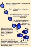
Activation of B cells occurs when a BCR (antibody) encounters and ligates its cognate antigen. Naïve B cells each have one of millions of distinct surface antigen-specific receptors, yet have not encountered their specific, cognate antigen. With a life-span of only a few days, many B cells die without ever encountering their cognate antigen. In most cases, B-cell activation is dependent upon costimulation by an activated helper T cell that has itself been activated by the same antigen. (click images to enlarge)

Unlike T cells, B cells are coated in immunoglobulin receptors and are able to recognize intact antigen, which they engulf, digest, and subsequently present in complex with surface MHC class II molecules. The MHC-peptide complex binds CD4 + helper T cells (Th), inducing secretion of cytokines that stimulate B cell proliferation and their differentiation into plasma cells, which secrete specific antibodies that bind with the cognate antigen. These antigen-antibody complexes are subsequently cleared by liver and spleen cells and the classical complement cascade.

Activation of T cells requires (1) TCR engagement, which ensures antigen specificity and MHC restriction of the response. However, synergistic signaling by (2) costimulatory molecules is also necessary to sustain and integrate TCR signaling to stimulate optimal T cell proliferation and differentiation.

Delivery of first signal (TCR engagement) in the absence of costimulation by a second signal(s) results in apoptosis or anergy. Anergic T cells neither produce IL-2 nor proliferate upon restimulation. This requirement of naïve T cell activation for delivery of both antigen-specific and costimulatory signals implies that only professional antigen presenting cells can initiate T cell responses.

Activation-regulatory mechanisms:
● increasing TCR avidity (adhesion molecules)
● enhancing recruitment of tyrosine kinases to the TCR complex coreceptors (CD4 and CD8)
● costimulation involving reciprocal and sequential signals between cells

Negative regulators of costimulation include receptors that bind B7 family members:
CTLA-4
● PD-1

Molecules involved in costimulation include:
1. Disulfide-linked homodimers that bind to distinct members of the B7 family of surface proteins
---CD28
---● ICOS (inducible costimulator) molecules
2. Members of the TNF receptor (TNFR) family
---CD40, the major B cell costimulatory molecule
---CD30
---CD27
---● OX-40
---● 4-1BB

The CD28 receptor is involved in the best characterized costimulatory pathway. CD28 is the primary costimulatory molecule for naïve T cells, although CD4+ helper T cells are more dependent than are CD8+ killer T cells on CD28 costimulation. CD28 binds the CD80 (B7-1) and CD86 (B7-2) ligands that are expressed on antigen presenting cells (APCs). CD28 costimulation increases T cell responses in naïve cells by increasing cytokine (mainly IL-2) production, which results from an increase in both cytokine gene transcription and mRNA stabilization.

CD28 signaling involves the activation of the small Rho family GTPases Rac and CDC42, which activate p21-activated kinase. This may link them to the mitogen-activated protein kinase cascades and the subsequent induction of IL-2 synthesis. Rac and CDC42 are also important in CD28-mediated cytoskeletal rearrangements, through the action of the Wiscott-Aldrich syndrome protein (WASP).

CD28 costimulation increases the activity of nuclear transcription factors of the Rel/NFkB family, whose members bind the CD28-responsive element (CD28RE) present in several cytokine gene promoters.

CD28 triggering reduces the number of engaged TCRs necessary to induce cytokine production and cell proliferation. This threshold reduction for T-cell activation is attributed to CD28-induced recruitment of lipid rafts to the immunological synapse, which promotes recruitment of raft-associated kinase and adapter molecules.

activation B : activation T : anergy : CD28 receptor : CD28RE : CDC42 : costimulatory molecules : first/second signals : helper T cell : IL-2 : MHC class II : negative regulators : plasma cells : Rac : regulatory mechanisms : Rel-NFkB : Rho GTPases : TCR engagement : TCR threshold reduction : WASP

Tables  Fc receptors  Immune Cytokines  Immunoglobulins

Top

tags

Labels: , , , , , , , , ,

| 0 Guide-Glossary

MHC

The major histocompatibility complex (MHC) encodes molecules displayed on cell surfaces, where they participate in lymphocyte recognition and antigen presentation.

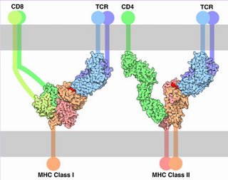
Left - click to enlarge - the presented antigen of the MHC-peptide complex is indicated by red. (Image source Molecule of Month: Illustration by David S. Goodsell of The Scripps Research Institute. Original work of the US Federal Government - public domain.)

The TCR and MHC-peptide complex make primary contact, then CD molecules bind to other portions of the MHC.

Almost ubiquitous, MHC class I interacts with TCRs and CD8 on cytotoxic T cells (CTCs, 'suppressor' T cells, regulator T cells). MHC class II is expressed primarily on cells that interact with pathogenic peptides. MHC II interacts with TCRs and with CD4 on helper T cells and cells that stimulate the immune system.

Proteins encoded by MHC genes identify the molecule as non-self or self. Non-self antigens – partly digested by lysosomes of leukocytes (monocytes or neutrophils) or displayed intact – are carried on the APC surface by class II histocompatibility molecules. Proteins of tumor cells or foreign invaders such as bacteria and viruses are carried by class I histocompatibility molecules. Non-self antigens provoke the immune response.

● Class I – encoded in BCA region – occur on almost every nucleated cell of the body, and are heterodimeric peptide binding proteins, antigen processing molecules such as TAP and Tapasin
● Class II – encoded in D region – occur only on specialized cell types, and are heterodimeric peptide binding proteins, proteins such as MHC II DM, MHC II DQ, and MHC II DP that that modulate peptide loading in the lysosomal compartment
● Class III – encoded between BCA and D regions – complement components such as C2, C4, factor B, and some that encode cytokines (e.g., TNF-α).

The Class I and Class II MHC molecules belong to molecules of the immunoglobulin supergene family, including immunoglobulins, T-cell receptors, CD4, and CD8. The major histocompatibility complex is encoded by several genes located on human chromosome 6 – class I molecules are encoded by the BCA region, while class II molecules are encoded by the D region. Sequences in the region between the BCA and D segments encodes class III molecules, which include some complement components and cytokines.

Human class II molecules are designated HLA-D, for human leukocyte antigen D, and the genes encoding them are also located in the major histocompatibility complex (MHC)

Tables  Fc receptors  Immune Cytokines  Immunoglobulins

æ Archaea & Eubacteria ¤ Cancer Cell Biology ~ Chemistry of Life  Diagrams & TablesEnzymesEvo Devo Molecular Biology φ Molecules ō Organics ››› Pathways ▫▫ Virus

Labels: , , , , ,

| 0 Guide-Glossary

. . . since 10/06/06
Google