Immunology

anergy

Anergy (immunologic tolerance) refers to the failure to mount a full immune response against a target.

Anergy toward self-targets operates as one self-tolerance mechanism to control the autoreactive cells found in autoimmunity. Clonal deletion in which lymphocytes are killed if they recognize a self-antigen during their maturation in the thymus gland or bone marrow is a major mechanism for the prevention of autoimmunity. However, not all human self-antigens are expressed in the central lymphoid organs where the lymphocytes are developing. Thus, self-tolerance to an individual's own antigens must also depend on mechanisms such as clonal anergy. Theoretically, recognition of a self-antigen eliminates the proliferative capacity of autoreactive lymphocytes in the peripheral immune system. Another process, immunoregulation, utilizes regulatory T cells that weaken harmful or inappropriate lymphocyte responses.

In B cell anergy, self-reactive B cells persist in the periphery yet remain unresponsive to immunogen. Research findings indicate that continuous binding of antigen and subsequent receptor signaling are essential for the maintenance of anergy.[n]

T cell anergy is induced when TCR stimulation "freezes" T cell responses until they receive an adequate subsequent antigenic signal from an antigen-presenting cell. Such APC signals can rescue T cells from anergy, stimulating them to produce the lymphokines necessary for the growth of additional T cells.

During a productive immune response, CD4+ T cells respond to effective signals by producing interleukin 2 (IL-2) and by proliferating. Effective signals stimulate require both ligation of TCRs with cognate antigens presented by class II MHC molecules on the surface of APCs and activation of costimulatory receptors, such as CD28, which recognize ligands such as B7 proteins expressed on the surface of APCs.

When T cells receive stimulus only TCR signals in the absence of engagement of costimulatory receptors, they enter a state of anergic unresponsiveness characterized by an inability to produce IL-2 or to proliferate upon re-stimulation. Such anergic T cells show a profound block in Ras/MAPK pathway that prevents activation of the AP-1 family of transcription factors (Fos/Jun).

GRAIL (gene related to anergy in lymphocytes) is GRAIL is an E3 ubiquitin ligase that is necessary for the induction of CD4+ T cell anergy in vivo. It is upregulated in naturally occurring (thymically derived) CD4+ and CD25+ cells [a] and anergized T cells [1]. Both GRAIL and Foxp3 are genotypic marker for CD25+ Treg cells. T cell activation appears to be controlled by Foxp3 through transcriptional regulation of early growth response (Egr) genes Egr-2 and Egr-3, and E3 ubiquitin (Ub) ligase genes Cblb [?], Itch [?] and GRAIL, subsequently affecting degradation of two key signaling proteins, PLCgamma1 and PKC-theta. [a]

It is believed that GRAIL could induce anergy through ubiquitylation of membrane-associated targets required for T-cell activation. It has been demonstrated that two isoforms of otubain-1, in conjunction with the deubiquitylating enzyme USP8, produce opposing effects on the expression and function of GRAIL in the induction of anergy.[2] GRAIL is differentially expressed in naturally occurring and peripherally induced CD25+ Treg cells where the expression of GRAIL has been suggested is linked to their functional "regulatory" activity.

Tables  Complement Receptors  Cytokines  Fc receptors  Immune Cytokines  Immunoglobulins  Interferons  Scavenger Receptors  Toll-like Receptors

tags

Labels: , , , , , , , , , , , , , , , , ,

| 0 Guide-Glossary

costimulation

Costimulation involves ligand-receptor interactions at the surfaces of a responder lymphocyte and an "accessory" cell – APCs for activation of T cells, and helper T cells for activation of B cells.

activation B : activation T : anergy : CD28 receptor : CD28RE : CDC42 : costimulatory molecules : first/second signals : helper T cell : IL-2 : MAPK cascade : MHC class II : negative regulators : Rac : regulatory mechanisms : Rel-NFkB : Rho GTPases : TCR engagement : TCR threshold reduction : transcription factors : WASP

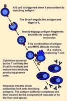
Activation of B cells occurs when a BCR (antibody) encounters and ligates its cognate antigen. Naïve B cells each have one of millions of distinct surface antigen-specific receptors, yet have not encountered their specific, cognate antigen. With a life-span of only a few days, many B cells die without ever encountering their cognate antigen. In most cases, B-cell activation is dependent upon costimulation by an activated helper T cell that has itself been activated by the same antigen. (click images to enlarge)

Unlike T cells, B cells are coated in immunoglobulin receptors and are able to recognize intact antigen, which they engulf, digest, and subsequently present in complex with surface MHC class II molecules. The MHC-peptide complex binds CD4 + helper T cells (Th), inducing secretion of cytokines that stimulate B cell proliferation and their differentiation into plasma cells, which secrete specific antibodies that bind with the cognate antigen. These antigen-antibody complexes are subsequently cleared by liver and spleen cells and the classical complement cascade.

Activation of T cells requires (1) TCR engagement, which ensures antigen specificity and MHC restriction of the response. However, synergistic signaling by (2) costimulatory molecules is also necessary to sustain and integrate TCR signaling to stimulate optimal T cell proliferation and differentiation.

Delivery of first signal (TCR engagement) in the absence of costimulation by a second signal(s) results in apoptosis or anergy. Anergic T cells neither produce IL-2 nor proliferate upon restimulation. This requirement of naïve T cell activation for delivery of both antigen-specific and costimulatory signals implies that only professional antigen presenting cells can initiate T cell responses.

Activation-regulatory mechanisms:
● increasing TCR avidity (adhesion molecules)
● enhancing recruitment of tyrosine kinases to the TCR complex coreceptors (CD4 and CD8)
● costimulation involving reciprocal and sequential signals between cells

Negative regulators of costimulation include receptors that bind B7 family members:
CTLA-4
● PD-1

Molecules involved in costimulation include:
1. Disulfide-linked homodimers that bind to distinct members of the B7 family of surface proteins
---CD28
---● ICOS (inducible costimulator) molecules
2. Members of the TNF receptor (TNFR) family
---CD40, the major B cell costimulatory molecule
---CD30
---CD27
---● OX-40
---● 4-1BB

The CD28 receptor is involved in the best characterized costimulatory pathway. CD28 is the primary costimulatory molecule for naïve T cells, although CD4+ helper T cells are more dependent than are CD8+ killer T cells on CD28 costimulation. CD28 binds the CD80 (B7-1) and CD86 (B7-2) ligands that are expressed on antigen presenting cells (APCs). CD28 costimulation increases T cell responses in naïve cells by increasing cytokine (mainly IL-2) production, which results from an increase in both cytokine gene transcription and mRNA stabilization.

CD28 signaling involves the activation of the small Rho family GTPases Rac and CDC42, which activate p21-activated kinase. This may link them to the mitogen-activated protein kinase cascades and the subsequent induction of IL-2 synthesis. Rac and CDC42 are also important in CD28-mediated cytoskeletal rearrangements, through the action of the Wiscott-Aldrich syndrome protein (WASP).

CD28 costimulation increases the activity of nuclear transcription factors of the Rel/NFkB family, whose members bind the CD28-responsive element (CD28RE) present in several cytokine gene promoters.

CD28 triggering reduces the number of engaged TCRs necessary to induce cytokine production and cell proliferation. This threshold reduction for T-cell activation is attributed to CD28-induced recruitment of lipid rafts to the immunological synapse, which promotes recruitment of raft-associated kinase and adapter molecules.

activation B : activation T : anergy : CD28 receptor : CD28RE : CDC42 : costimulatory molecules : first/second signals : helper T cell : IL-2 : MHC class II : negative regulators : plasma cells : Rac : regulatory mechanisms : Rel-NFkB : Rho GTPases : TCR engagement : TCR threshold reduction : WASP

Tables  Fc receptors  Immune Cytokines  Immunoglobulins

Top

tags

Labels: , , , , , , , , ,

| 0 Guide-Glossary

T cells

T cells or T lymphocytes are specialized lymphocytes distinguished by their TCR surface receptors.

activation T reg : APC : apoptosis : B7-1 : B7-2 : CD8 + : CD28 : CD80 : CD86 : costimulation : CTL : cytotoxic T lymphocytes : Fas-FasL : γδ T cells : granulysin : granzyme : helper T cell : IL-2 : interleukin-2 : natural killer T cells : NKT : perforin : pore : self-protection : serine protease : Tc : Th : Treg : Treg activation/action : T cell types : VDJ recombination

'T' lymphocytes initially develop in the thymus, and differentiate peripherally into several functional subsets of cells:
cytotoxic T cellsCTL, TcCD8 +
helper T cells, effector T cells – ThCD4 +
regulatory T cellsTreg (formerly termed 'suppressor' T cells)
natural killer T cellsNKT (different than NK cells)
γδ T cells

Cytotoxic T lymphocytes (CTLs) or killer T cells express antigen-specific, VDJ recombination-generated TCRs plus glycoprotein CD8 on their surfaces. The TCR recognizes specific antigenic proteins that are fragmented into peptides and then are presented in complex with MHC class I molecules (MHC-peptide complex - red).

APCs provide synergistic signaling by costimulatory molecules. APC signaling is necessary to sustain and integrate TCR signaling and to stimulate optimal T cell proliferation and differentiation.

The binding of TCR•antigen•CD8•MHC-I holds the activated CTL (Tc) in proximity to the damaged/infected target cell. Once activated, the Tc cell undergoes clonal expansion with the assistance of a cytokine, interleukin-2 (IL-2) that acts as a growth and differentiation factor for T cells. (Table  Immune Cytokines)

CD28 binds the CD80 (B7-1) and CD86 (B7-2) ligands that are expressed on antigen presenting cells (APCs). When exposed to infected/dysfunctional somatic cells, Tc cells release perforin, which form pores in the target cell's plasma membrane. Also released from Tcs are granulysin and the serine protease granzyme, which transit the perforin pore and induce apoptosis of the target cell.

A second cytolytic mechanism is mediated by the interaction of activated T cell surface FasL with TNFR family Fas receptors (TNFRSF6, Apo_1, CD95) expressed on the surface of the target cell.

Helper T cells (Th), or effector T cells circulate throughout the body where they interface with MHC class II protein on other cells, determining whether the MHC class II is presenting 'self' or 'non-self protein' (antigen). Th cells direct antibody class switching in B lymphocytes.

When the helper T cell is activated by contact with antigen, it enters the cell cycle in addition to producing lymphokines and chemokines. Th cells orchestrate activation and growth of cytotoxic T cells, and maximize the bactericidal activity of phagocytes (macrophages).

Helper T cells mostly carry the CD4 surface protein, though a few carry CD8. The CD4 receptor triggers targetting by HIV, which determines the crippling effect of HIV on the immune system.

Regulatory cells (Treg)
Regulatory T cells were formerly called suppressor T cells because they suppress immune responses. Negative regulatory functions have been ascribed to several T cell types:
CD4+CD25+
CD4+CD25–
interleukin-10-producing TR1 cells
transforming growth factor-ß-secreting TH3 'regulatory' cells
[v]

Treg cells express the αβ TCR chains and are activated only when the TCR binds to its cognate peptide-class II MHC molecule and receives costimulation from B7 molecules (CD80, CD86) on the antigen-presenting cell. Once activated, Treg secrete interleukin 9 (IL-9), interleukin 10 (IL-10), +/- transforming growth factor-beta (TGF-β), and these lymphokines inhibit Th1 assistance of cell-mediated immunity and inflammation, and
Th2 assistance of antibody production, and may inhibit the action of CD8+ CTLs.

Because the antigenic peptides recognized by Treg TCRs are typically self-peptides, the chief function of Treg cells is provision of self-protection by preventing other T cells from attacking self components, thus protecting against autoimmunity.

CD25, or IL-2 receptor alpha chain, or IL-2R, or Tac antigen is a 55kDa glycoprotein whose transcription is regulated by Stat5, Elf-1, HGG-I(Y), GATA, and Tax from HTLV-1. CD25 is expressed on PHA-stimulated T cells, B cells stimulated with anti-IgM antibody, monocytes/macrophages stimulated with LPS, and highly expressed on HTLV-I-transformed T cell lines. NCBI LocusLink Record: 3559

Natural killer T cells – NKT cells are activated by group 2, CD1d molecules to secrete interferon-γ (IFNγ) and IL-4, which act as Th1 and Th2 cytokines.

γδ T cells carry TCRs encoded by different gene segments than those of αβ T cells. CD27 is cell-surface marker for T-cell progenitors that are beginning to undergo commitment to the αβ or γδ T-cell lineages, and newly emerging αβ and γδ T-lineage cells display early differences in dependence on Notch–Delta signaling and display distinct patterns of gene expression.[npg]

Presentation of peptide antigens for activation of naïve T cells does not reside solely in dendritic cells. A population of γδ T cells can efficiently present peptide antigens to αβT cells, which comprise the predominant mammalian T cell population. Thus, γδ T cells function to bridge the innate and adaptive immune responses.

γδ T cells can form unique memory subsets with distinct adhesion and chemotactic capabilities, determining the tissue homing capabilities of cells. γδ T cells are enriched in areas of inflammation. Human γδ T cells express ligands for E- and P-selectins, so they display specialized homing to epithelial surfaces. The memory γδ T cells express chemokine family receptors not expressed by αβ T cells.

Human γδ T cells that express Vγ2Vδ2 TCRs can recognize nonpeptide antigens and Non-Hodgkin's B cell lymphomas. In particular, recognition of prenyl pyrophosphates is performed by this extracellular presentation pathway, which is distinct from MHC and CD1 presentation, and which is important in many different bacterial and parasitic infections. However, γδ T cells of the major tissue subset recognize self and foreign nonpeptide antigens presented by CD1. These nonpeptide antigens are lipids and glycolipids presented by CD1 proteins, which are MHC-like-molecules specialized for the presentation of lipids.[s].


▲ф ф antibodies ф antigen : APC ф APCs : apoptosisapoptosis : B7-1 : B7-2 ф CD : CD8 + : CD28 : CD80 : CD86cell growthcell membranescellular fate : costimulation ф costimulation : CTL ~ cytokines ф cytolysis : cytotoxic T lymphocytesdifferentiation : Fas-FasL : γδ T cells : granulysin § granulysin : granzyme : helper T cell ф helper T cell : IL-2 ~ immunoglobulins : interleukin-2 ф immune cytokines  Immune Cytokines : killer T cells ф killer T cells ф lymphocytes ф lymphokines ф MHC : natural killer T cells : NKT : perforin : pore ф receptors : serine protease ф signaling ф surface receptors: Tc : Th : Treg : T cell types ф thymus : VDJ recombination ф VDJ recombination ▲ф

Tables  Cell Adhesion Molecules  Cell signaling  Complement Receptors  Cytokines  Fc receptors  Immunoglobulins  Interferons  Receptor Tyrosine Kinases (RTKs)  Receptor Signal Transduction  Scavenger Receptors  Second Messengers  Toll-like Receptors
Top

æ Archaea & Eubacteria ¤ Cancer Cell Biology ~ Chemistry of Life  Diagrams & TablesEnzymesEvo Devo Molecular Biology φ Molecules ō Organics ››› Pathways ▫▫ Virus

Labels: , , , , , , , ,

| 0 Guide-Glossary

. . . since 10/06/06
Google