Immunology

anergy

Anergy (immunologic tolerance) refers to the failure to mount a full immune response against a target.

Anergy toward self-targets operates as one self-tolerance mechanism to control the autoreactive cells found in autoimmunity. Clonal deletion in which lymphocytes are killed if they recognize a self-antigen during their maturation in the thymus gland or bone marrow is a major mechanism for the prevention of autoimmunity. However, not all human self-antigens are expressed in the central lymphoid organs where the lymphocytes are developing. Thus, self-tolerance to an individual's own antigens must also depend on mechanisms such as clonal anergy. Theoretically, recognition of a self-antigen eliminates the proliferative capacity of autoreactive lymphocytes in the peripheral immune system. Another process, immunoregulation, utilizes regulatory T cells that weaken harmful or inappropriate lymphocyte responses.

In B cell anergy, self-reactive B cells persist in the periphery yet remain unresponsive to immunogen. Research findings indicate that continuous binding of antigen and subsequent receptor signaling are essential for the maintenance of anergy.[n]

T cell anergy is induced when TCR stimulation "freezes" T cell responses until they receive an adequate subsequent antigenic signal from an antigen-presenting cell. Such APC signals can rescue T cells from anergy, stimulating them to produce the lymphokines necessary for the growth of additional T cells.

During a productive immune response, CD4+ T cells respond to effective signals by producing interleukin 2 (IL-2) and by proliferating. Effective signals stimulate require both ligation of TCRs with cognate antigens presented by class II MHC molecules on the surface of APCs and activation of costimulatory receptors, such as CD28, which recognize ligands such as B7 proteins expressed on the surface of APCs.

When T cells receive stimulus only TCR signals in the absence of engagement of costimulatory receptors, they enter a state of anergic unresponsiveness characterized by an inability to produce IL-2 or to proliferate upon re-stimulation. Such anergic T cells show a profound block in Ras/MAPK pathway that prevents activation of the AP-1 family of transcription factors (Fos/Jun).

GRAIL (gene related to anergy in lymphocytes) is GRAIL is an E3 ubiquitin ligase that is necessary for the induction of CD4+ T cell anergy in vivo. It is upregulated in naturally occurring (thymically derived) CD4+ and CD25+ cells [a] and anergized T cells [1]. Both GRAIL and Foxp3 are genotypic marker for CD25+ Treg cells. T cell activation appears to be controlled by Foxp3 through transcriptional regulation of early growth response (Egr) genes Egr-2 and Egr-3, and E3 ubiquitin (Ub) ligase genes Cblb [?], Itch [?] and GRAIL, subsequently affecting degradation of two key signaling proteins, PLCgamma1 and PKC-theta. [a]

It is believed that GRAIL could induce anergy through ubiquitylation of membrane-associated targets required for T-cell activation. It has been demonstrated that two isoforms of otubain-1, in conjunction with the deubiquitylating enzyme USP8, produce opposing effects on the expression and function of GRAIL in the induction of anergy.[2] GRAIL is differentially expressed in naturally occurring and peripherally induced CD25+ Treg cells where the expression of GRAIL has been suggested is linked to their functional "regulatory" activity.

Tables  Complement Receptors  Cytokines  Fc receptors  Immune Cytokines  Immunoglobulins  Interferons  Scavenger Receptors  Toll-like Receptors

tags

Labels: , , , , , , , , , , , , , , , , ,

| 0 Guide-Glossary

B cells

B cells are lymphocytes (WBCs) that participate in humoral immunity by producing antibodies in response to antigen stimulation.

activation : B-1 : B-2 : BCRs : CDRs : granzymes : helper T cells : life-span B cells : lymphopoiesis : memory B : naïve B cells : NK cells : NK receptors : NK cells attack viral infected cells : perforin : plasma B : stimulation : surface-immunoglobulins : surface receptors : VDJ recombination

Surface membrane-associated immunoglobulins (IgD and IgM) act as B cell receptors (BCRs), and the enormous variety of antigen recognition sites is attributable to VDJ recombination (alternative splicing) of peptide sequences encoded by V, D, and J genes. The variable region of immunoglobulins includes the recognition sites or complementarity determining regions (CDRs).
Lymphopoiesis, which takes place in the bone marrow of almost all mammals, produces small lymphocytes, large granular lymphocytes (NK) cells, B lymphocytes (precursors of plasma cells, T lymphocytes, and lymphoid dendritic cell. Recognition of self during lymphopoiesis permits anergy (suppression of self-attack).

Naïve B cells each have one of millions of distinct surface antigen-specific receptors, yet have not encountered their specific, cognate antigen. With a life-span of only a few days, many B cells die without ever encountering their cognate antigen. Naïve B cells are activated when the BCR binds to its cognate antigen. This antigen-Ig binding must be coupled with a signal from a helper T cell in order to activate the B cell.

Once activated, B lymphocytes:
● differentiate into one of the B cell types (directly or through intermediate, germinal center reactions)
● plasma cells produce antibodies against the antigenic stimulus, or memory cells are primed for subsequent activation by the antigen

Types of B cell:
B-1
B-2
Plasma B cells
Memory B cells

After newly formed B cells exit generative sites in fetal liver or adult bone marrow they undergo selection events that may involve interactions with self or with external antigens. Selective events can influence the phenotype and functional characteristics of B cells. B cell receptor-mediated events also influence lymphoid organs localization as marginal zone B cells in the spleen, as follicular (B-2 cells), as well as B-1 cells in the peritoneal and pleural cavities. [] fluorescence micrograph spleen, fm high power in which T cells form periarteriolar lymphocyte sheath (PALS) (red) and B-2 cell follicles (green) []

B-1 cells are the first B cells produced in the fetus, and in adults are located primarily in the peritoneal and pleural cavities. B1 cells are believed to operate in the innate response to infection by viruses and bacteria, and usually show preferential responses to T cell-independent antigens. The diversity of B-1 lymphocytes is attributed to their recombinatorial recombination, in which there is a preferential recombination between D-proximal VH gene segments. B-1 lymphocytes express (polyspecific) IgM in greater quantities than they express IgG, and the ability of B1 cells to respond to isotype switch commitment factors such as interleukin-4 may be secondary to their production of IgM. B-1 cells express CD5, which binds to CD72 to mediate B cell-B cell interactions.

B-2 cells are conventional B lymphocytes that are produced postnatally (unlike fetal B-1 cells) and are replaced from the bone marrow.

Plasma B lymphocytes are committed to production of copious amounts of monoclonal antibodies.

Memory B lymphocytes are long-lived, stimulated B lymphocytes that are primed for rapid response to a repeated exposure of the priming antigen. Memory B cells are generated in lymphoid tissue after B cell activation/proliferation and reside in the bone marrow, lymph nodes, and spleen. High affinity surface immunoglobulins enable their activation by lower levels of cognate antigen than are naïve B cells.

NK cells are differentiated from killer T cells. NK, natural killer cells constitute a corps of circulating lymphocytes that are constitutively specialized to attack cancerous cells and virus infected cells. Preprogramming for target recognition, coupled with the absense of need for backup by a clone of identical cells, renders NK cells capable of rapid (innate) response to pathogens. NK attack involves the exocytosis of cytoplasmic granules containing perforin and granzymes. Perforin forms pores in the plasma membrane of attacked cells through which serine-protease granzymes enter, cleaving caspase precursors and triggering apoptosis.

Individuals inherit multiple, polymorphic genes for NK receptors, so the assemblage of NK receptors differs between individuals. NK cells carry two forms of surface receptors:
● killer inhibitory receptors (KIRs) transmit an inhibitory signal when they encounter class I MHC molecules on a cell surface. (By contrast, T cells only recognize antigens that are presented by a MHC molecule.)
● activating receptors, which activate the NK cell upon binding to a target cell

Viral infection often causes suppression of MHC expresion, leading to a reduction of inhibition of NKs by its killer inhibitory receptors. This double negative renders the virus infected cell a target for killing by NK cells.

"About 85% of peripheral B cells are phenotypically mature and display first-order exponential kinetics defined by a half-life of 5-6 weeks, whilst the remainder are short-lived with a life span of several days."[s]

[] tem plasma cell [] micrograph macrophage surrounded by normal plasma cells [] micrograph macrophage & plasma cells []

activation : BCRs : CDRs : helper T cells : life-span B cells : lymphopoiesis : naïve B cells : surface-immunoglobulins : surface receptors : VDJ recombination

Tables  Fc receptors  Immune Cytokines  Immunoglobulins  Cell Adhesion Molecules  Cell signaling  Receptor Tyrosine Kinases (RTKs)  Receptor Signal Transduction  Second Messengers 

Top

tags

Labels: , , , , , ,

| 0 Guide-Glossary

costimulation

Costimulation involves ligand-receptor interactions at the surfaces of a responder lymphocyte and an "accessory" cell – APCs for activation of T cells, and helper T cells for activation of B cells.

activation B : activation T : anergy : CD28 receptor : CD28RE : CDC42 : costimulatory molecules : first/second signals : helper T cell : IL-2 : MAPK cascade : MHC class II : negative regulators : Rac : regulatory mechanisms : Rel-NFkB : Rho GTPases : TCR engagement : TCR threshold reduction : transcription factors : WASP

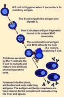
Activation of B cells occurs when a BCR (antibody) encounters and ligates its cognate antigen. Naïve B cells each have one of millions of distinct surface antigen-specific receptors, yet have not encountered their specific, cognate antigen. With a life-span of only a few days, many B cells die without ever encountering their cognate antigen. In most cases, B-cell activation is dependent upon costimulation by an activated helper T cell that has itself been activated by the same antigen. (click images to enlarge)

Unlike T cells, B cells are coated in immunoglobulin receptors and are able to recognize intact antigen, which they engulf, digest, and subsequently present in complex with surface MHC class II molecules. The MHC-peptide complex binds CD4 + helper T cells (Th), inducing secretion of cytokines that stimulate B cell proliferation and their differentiation into plasma cells, which secrete specific antibodies that bind with the cognate antigen. These antigen-antibody complexes are subsequently cleared by liver and spleen cells and the classical complement cascade.

Activation of T cells requires (1) TCR engagement, which ensures antigen specificity and MHC restriction of the response. However, synergistic signaling by (2) costimulatory molecules is also necessary to sustain and integrate TCR signaling to stimulate optimal T cell proliferation and differentiation.

Delivery of first signal (TCR engagement) in the absence of costimulation by a second signal(s) results in apoptosis or anergy. Anergic T cells neither produce IL-2 nor proliferate upon restimulation. This requirement of naïve T cell activation for delivery of both antigen-specific and costimulatory signals implies that only professional antigen presenting cells can initiate T cell responses.

Activation-regulatory mechanisms:
● increasing TCR avidity (adhesion molecules)
● enhancing recruitment of tyrosine kinases to the TCR complex coreceptors (CD4 and CD8)
● costimulation involving reciprocal and sequential signals between cells

Negative regulators of costimulation include receptors that bind B7 family members:
CTLA-4
● PD-1

Molecules involved in costimulation include:
1. Disulfide-linked homodimers that bind to distinct members of the B7 family of surface proteins
---CD28
---● ICOS (inducible costimulator) molecules
2. Members of the TNF receptor (TNFR) family
---CD40, the major B cell costimulatory molecule
---CD30
---CD27
---● OX-40
---● 4-1BB

The CD28 receptor is involved in the best characterized costimulatory pathway. CD28 is the primary costimulatory molecule for naïve T cells, although CD4+ helper T cells are more dependent than are CD8+ killer T cells on CD28 costimulation. CD28 binds the CD80 (B7-1) and CD86 (B7-2) ligands that are expressed on antigen presenting cells (APCs). CD28 costimulation increases T cell responses in naïve cells by increasing cytokine (mainly IL-2) production, which results from an increase in both cytokine gene transcription and mRNA stabilization.

CD28 signaling involves the activation of the small Rho family GTPases Rac and CDC42, which activate p21-activated kinase. This may link them to the mitogen-activated protein kinase cascades and the subsequent induction of IL-2 synthesis. Rac and CDC42 are also important in CD28-mediated cytoskeletal rearrangements, through the action of the Wiscott-Aldrich syndrome protein (WASP).

CD28 costimulation increases the activity of nuclear transcription factors of the Rel/NFkB family, whose members bind the CD28-responsive element (CD28RE) present in several cytokine gene promoters.

CD28 triggering reduces the number of engaged TCRs necessary to induce cytokine production and cell proliferation. This threshold reduction for T-cell activation is attributed to CD28-induced recruitment of lipid rafts to the immunological synapse, which promotes recruitment of raft-associated kinase and adapter molecules.

activation B : activation T : anergy : CD28 receptor : CD28RE : CDC42 : costimulatory molecules : first/second signals : helper T cell : IL-2 : MHC class II : negative regulators : plasma cells : Rac : regulatory mechanisms : Rel-NFkB : Rho GTPases : TCR engagement : TCR threshold reduction : WASP

Tables  Fc receptors  Immune Cytokines  Immunoglobulins

Top

tags

Labels: , , , , , , , , ,

| 0 Guide-Glossary

. . . since 10/06/06
Google