Immunology

cellular response

Cellular responses to invading pathogens utilize phagocytic and cytotoxic cells of the innate and adaptive immune responses.

The immune system is intimately connected with the hematologic system since white blood cells (leukocytes, including B- and T-lymphocytes) are key players in the lymphoid system.
Cellular participants in the immune and inflammatory responses include :
phagocytic cells (dendritic cells, monocytes and macrophages, and granulocytes)
antigen presenting cells (dendritic cells, macrophages, B lymphocytes, helper T cells, γδ T cells)
antibody producing cells (plasma cells)
cytotoxic cells (CTL, NK)
● regulatory cells (APCs, helper T cells, regulatory T cells)
● cells-in-waiting (memory B cells, monocytes)
● chemical releasing cells (basophils, eosinophils, neutrophils; mast cells - histamine, cytokines; hepatocytes - complement proteins)

Innate responses solely comprise cellular immune responses employ phagocytic cells that are circulating or tissue emplaced – granulocytes, monocytes, dendritic cells, macrophages, natural killer T cells, and B lymphocytes. The innate response induces (triggers) the adaptive system, the cellular component of which relies upon activated macrophages, T-lymphocytescytotoxic T lymphocytes (killer T cells).

Tables  Fc receptors  Immune Cytokines  Immunoglobulins

tags

Labels: , , , , , , , , , ,

| 0 Guide-Glossary

complement system

The complement system comprises an assembly of liver-manufactured, soluble and cell-bound proteins that participate in innate and adaptive immunity. Activation of the complement cascade by protease cleavage leads to chemotaxis (C5a), inflammation and increased capillary permeability (C3a, C5a), opsonization (C3b), and cytolysis.

activation : alternative pathway : amplification by C3 : anaphylatoxins C3a, C5a : antigen uptake : C1 : C2 : C3 : C4 : C5 : C6 : C7 : C8 : C9 : C1INH : CD59 : chemotaxis : classical pathway : complement cascade : complement control proteins : convertases C3, C3/C5, C5 : disorders : evolution : Factor B : Factor H : Factor I : ficolins : immunoglobulins and complement activation : inhibitory proteins : lectin pathway : MBL -MAPS : mannose-binding lectin pathway : membrane attack complex : opsonin : pathways : perforin : phagocytosis : pore : proteases : regulation : serine proteases : sialic acid

Sequential activation of the protein components of the complement cascade upon cleavage by a protease, leads to each component's becoming, in its turn, a protease. Three pathways are involved in complement attack upon pathogens:
classical pathway
alternative pathway
mannose-binding lectin pathway (MBL -MAPS)

The classical pathway utilizes C1, which is activated by binding of an antibody to its cognate antigen.

Inactive C1 circulates as a serum molecular complex comprising 6 C1q molecules, 2 C1r molecules, and 2 C1s molecules. Constant regions in some immunoglobulins specifically bind C1q, activating C1r and C1s. The mu chains of IgM and some gamma chains of IgG contain specific binding sites, though IgM is far more effective than IgG.

Activated C1s is a serine protease that cleaves C4 and C2 into small inactive fragments (C4a, C2a) and larger active fragments, C4b and C2b. The active component C4b binds to the sugar moieties of surface glycoproteins and binds noncovalently to C2b, forming another serine protease C4b•C2b, which is called C3 convertase because it cleaves C3, releasing an active C3b opsonin fragment.

Macrophages and neutrophils possess receptors for C3b, so cells coated with C3b are targetted for phagocytosis (opsonization). The small C3a fragment is released into solution where it can bind to basophils and mast cells, triggering histamine release and, as an anaphylatoxin, potentially participating in anaphylaxis.

C3 amplifies the humoral response because of its abundance and its ability to auto-activate (as a C3 convertase). Breakdown of C3b generates an antigen-binding C3d fragment that enhances antigen uptake by dendritic cells and B cells .

Binding of C3b to C5 induces an allosteric change that exposes C3b•C5 to cleavage by C4b•C2b, which is now acting as C3/C5 convertase. The alternative pathway possesses a distinct C5 convertase, so the two pathways converge through C5.

Cleavage of C5 by the C3/C5 convertase releases:
anaphylotoxic C5a, which promotes chemotaxis of neutrophils
C5b, which complexes with one molecule of each of C6, C7, and C8. The resultant C5b•6•7•8 complex assists polymerization of as many as 18 C9 molecules to form a cytolysis-promoting pore (membrane attack complex, tem) through the plasma membrane of the target cell, which then suffers osmosis-induced cytolysis.

Another cytolytic mediator utilized by CTLs and NK cells is perforin, which is a 534 aa glycoprotein with sequence homology to the membrane attack component of complement C9. Like C9, perforin integrates into the target cell membrane, forming polyprotein pores up to 20nm in diameter comprising 12—18 perforin monomers, which breach membrane integrity and permit cytolytic cell death.

The alternative pathway is not activated by antigen-antibody binding, but instead relies upon spontaneous conversion of C3 to C3b, which is rapidly inactivated by its binding to inhibitory proteins and sialic acid on the cell's surface. Because bacteria and other foreign materials lack these inhibitory proteins and sialic acid, the C3b is not inactivated and it forms the C3b•Bb complex with Factor B. The C3b.Bb complex acts as a C3 convertase, forming C3b•Bb•C3b, which acts as a C5 convertase that can ititiate assembly of the membrane attack complex. C3b•Bb, acting as a C3 convertase, provides a positive feedback loop that amplifies production of C3.

The lectin pathway (MBL - MASP) is homologous to the classical pathway, but utilizes opsonin, mannan-binding lectin (MBL, MBP) and ficolins rather than C1q. Binding of mannan-binding lectin to mannose residues on the pathogen surface activates the MBL-associated serine proteases, MASP-1, MASP-2, MASP-3, which cleave C4 into C4b and C2 into C2b. As in the classical pathway, C4b and C2b bind to form the C4b•C2b C3 convertase. Ficolins are homologous to MBL and function through MASPs. Diversified ficolins are of particular importance in invertebrates, which lack the adaptive immune response that evolved some 500 million years ago in jawed vertebrates.

Several complement control proteins regulate activity of the complement system, including:
● C1 inhibitor (C1INH), which eliminates the proteolytic activity of activated C1r and C1s. Following C1 activation by antigen-antibody complexes, C1INH permits only a brief interval during which activated C1 can cleave C4 and C2.
● Factor I, which inactivates C3b
● Factor H, which removes Bb, thus interrupting the C3 convertase feedback loop within the alternative pathway
● CD59, which inhibits C9 polymerization during assembly of the membrane attack complex.

Dysregulation of the complement system manifests variously as immune complex disorders (C2 deficiency), susceptibility to bacterial infections (C3 deficiency), the autoimmune disorder SLE (early component or C2 deficiency), hereditary angioneurotic edema (HANE) (C1INH deficiency).

activation : alternative pathway : amplification by C3 : anaphylotoxins C3a, C5a ф antibodies ф antigen : antigen uptake : C1 : C2 : C3 : C4 : C5 : C6 : C7 : C8 : C9 : C1INH : CD59 : chemotaxis : classical pathway : complement cascade : complement control proteins : convertases C3, C3/C5, C5 : disorders : evolution : Factor B : Factor H : Factor I : ficolins : immunoglobulins and complement activation : inhibitory proteins : lectin pathway : MBL -MAPS : mannose-binding lectin pathway : membrane attack complex : opsonin : pathways : perforin : phagocytosis : pore : proteases : regulation : serine proteases : sialic acid

Tables  Fc receptors  Immune Cytokines  Immunoglobulins

Top

tags

Labels: , , , , , , , , , , ,

| 0 Guide-Glossary

costimulation

Costimulation involves ligand-receptor interactions at the surfaces of a responder lymphocyte and an "accessory" cell – APCs for activation of T cells, and helper T cells for activation of B cells.

activation B : activation T : anergy : CD28 receptor : CD28RE : CDC42 : costimulatory molecules : first/second signals : helper T cell : IL-2 : MAPK cascade : MHC class II : negative regulators : Rac : regulatory mechanisms : Rel-NFkB : Rho GTPases : TCR engagement : TCR threshold reduction : transcription factors : WASP

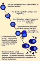
Activation of B cells occurs when a BCR (antibody) encounters and ligates its cognate antigen. Naïve B cells each have one of millions of distinct surface antigen-specific receptors, yet have not encountered their specific, cognate antigen. With a life-span of only a few days, many B cells die without ever encountering their cognate antigen. In most cases, B-cell activation is dependent upon costimulation by an activated helper T cell that has itself been activated by the same antigen. (click images to enlarge)

Unlike T cells, B cells are coated in immunoglobulin receptors and are able to recognize intact antigen, which they engulf, digest, and subsequently present in complex with surface MHC class II molecules. The MHC-peptide complex binds CD4 + helper T cells (Th), inducing secretion of cytokines that stimulate B cell proliferation and their differentiation into plasma cells, which secrete specific antibodies that bind with the cognate antigen. These antigen-antibody complexes are subsequently cleared by liver and spleen cells and the classical complement cascade.

Activation of T cells requires (1) TCR engagement, which ensures antigen specificity and MHC restriction of the response. However, synergistic signaling by (2) costimulatory molecules is also necessary to sustain and integrate TCR signaling to stimulate optimal T cell proliferation and differentiation.

Delivery of first signal (TCR engagement) in the absence of costimulation by a second signal(s) results in apoptosis or anergy. Anergic T cells neither produce IL-2 nor proliferate upon restimulation. This requirement of naïve T cell activation for delivery of both antigen-specific and costimulatory signals implies that only professional antigen presenting cells can initiate T cell responses.

Activation-regulatory mechanisms:
● increasing TCR avidity (adhesion molecules)
● enhancing recruitment of tyrosine kinases to the TCR complex coreceptors (CD4 and CD8)
● costimulation involving reciprocal and sequential signals between cells

Negative regulators of costimulation include receptors that bind B7 family members:
CTLA-4
● PD-1

Molecules involved in costimulation include:
1. Disulfide-linked homodimers that bind to distinct members of the B7 family of surface proteins
---CD28
---● ICOS (inducible costimulator) molecules
2. Members of the TNF receptor (TNFR) family
---CD40, the major B cell costimulatory molecule
---CD30
---CD27
---● OX-40
---● 4-1BB

The CD28 receptor is involved in the best characterized costimulatory pathway. CD28 is the primary costimulatory molecule for naïve T cells, although CD4+ helper T cells are more dependent than are CD8+ killer T cells on CD28 costimulation. CD28 binds the CD80 (B7-1) and CD86 (B7-2) ligands that are expressed on antigen presenting cells (APCs). CD28 costimulation increases T cell responses in naïve cells by increasing cytokine (mainly IL-2) production, which results from an increase in both cytokine gene transcription and mRNA stabilization.

CD28 signaling involves the activation of the small Rho family GTPases Rac and CDC42, which activate p21-activated kinase. This may link them to the mitogen-activated protein kinase cascades and the subsequent induction of IL-2 synthesis. Rac and CDC42 are also important in CD28-mediated cytoskeletal rearrangements, through the action of the Wiscott-Aldrich syndrome protein (WASP).

CD28 costimulation increases the activity of nuclear transcription factors of the Rel/NFkB family, whose members bind the CD28-responsive element (CD28RE) present in several cytokine gene promoters.

CD28 triggering reduces the number of engaged TCRs necessary to induce cytokine production and cell proliferation. This threshold reduction for T-cell activation is attributed to CD28-induced recruitment of lipid rafts to the immunological synapse, which promotes recruitment of raft-associated kinase and adapter molecules.

activation B : activation T : anergy : CD28 receptor : CD28RE : CDC42 : costimulatory molecules : first/second signals : helper T cell : IL-2 : MHC class II : negative regulators : plasma cells : Rac : regulatory mechanisms : Rel-NFkB : Rho GTPases : TCR engagement : TCR threshold reduction : WASP

Tables  Fc receptors  Immune Cytokines  Immunoglobulins

Top

tags

Labels: , , , , , , , , ,

| 0 Guide-Glossary

leukocytes

Leukocytes are white blood cells (WBCs) – left b=basophil, e=eosinophil, m=monocyte, n=neutrophil.

Leukocytes are subdivided according to presense or lack of granulation:

granulocytes generated by granulopoiesis :
--- basophilsmast cells
--- eosinophils
--- neutrophils

agranulocytes
-lymphocytes generated by lymphopoiesis
---B lymphocytesplasma cells
---T lymphocyteshelper T cells, killer T cells
--- ○ natural killer cells → lymphoid dendritic cells

- ○ agranulocytes generated by monocytopoiesis
---monocytesmacrophages
----------------.myeloid dendritic cells

Tables  Fc receptors  Immune Cytokines  Immunoglobulins

æ Archaea & Eubacteria ¤ Cancer Cell Biology ~ Chemistry of Life  Diagrams & TablesEnzymesEvo Devo Molecular Biology φ Molecules ō Organics ››› Pathways ▫▫ Virus

Labels: , , , , , , , , ,

| 0 Guide-Glossary

. . . since 10/06/06
Google