Immunology

basophils

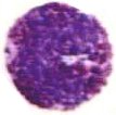
Basophils are granulocytes packed with granules that stain basic (purple with H&E). The nucleus is bilobed, and the metachromatic granules contain sulfated glycosaminoglycans as well as vasoactive compounds – histamine and proteoglycans. Lysosomal arylsulfatase is found in granules of developing basophils in bone marrow. Basophil granules are surrounded by a unit membrane and contain particles which are uniform in size across the same granule yet vary in size in different granules within the same cell. Some granules reveal a homogeneous texture and/or "myelin" figures. [s] Though basophils and mast cells share morphological features, the appearance of most basophil granules differs from the ultrastructure of human mast cell granules.

Activated basophils release proinflammatory histamine and proteoglycans from granules, and synthesize then secrete leukotrienes and cytokines (particularly IL-4). Histamine and IL-4, which is associated with production of IgE, are involved in allergic reactions.

Basophils are the least common granulocyte, representing about 0.5% to 1% of circulating leukocytes. A low basophil count combined with a low neutrophil count almost always portends leukemia.

Tables  Fc receptors  Immune Cytokines  Immunoglobulins .

tags

Labels: , , , , , , , , , , , , ,

| 0 Guide-Glossary

eosinophils

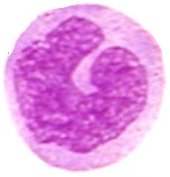
Eosinophils (acidophils) are granulocytes packed with granules that stain acidic (red with H&E). Eosinophils combat parasitic infection, and have diverse immune responsibilities.

Eosinophils play roles in:
● combatting viral infections (RNAses)
● allergic response
● fibrinolysis (following inflammation).
● pathogenesis of asthma
● combatting helminthic colonization, other parasites.

The acidophilic granules contain proteolytic enzymes that are toxic to parasite and elicit allergic symptoms in the host. The enzymes include histaminase, peroxidase, RNase, DNases, lipase, plasminogen, and Major Basic Protein. Eosinophils also secrete IL-5.

Eosinophils normally comprises about 2.3% of leukocytes, and elevations (eosinophilia) are associated with parasitic infestation, collagen vascular diseases (such as rheumatoid arthritis), malignancies such as Hodgkin's lymphoma, allergic skin conditions (such as exfoliative dermatitis), Addison's Disease, and hypersensitivity to drugs such as penicillin.

Tables  Fc receptors  Immune Cytokines  Immunoglobulins

tags

Labels: , ,

| 0 Guide-Glossary

granulocytes

Granulocytes are granulated leukocytes generated by granulopoiesis in the bone marrow. Because their nuclei vary in shape (usually 3-lobed) granulocytes are also termed polymorphonuclear leukocytes (PMN, PML). Usually, the term polymorphonuclear leukocyte is used to designate neutrophil granulocytes, which are the most abundant of the granulocytes.

The granules are named according to their staining characteristics with hematoxylin and eosin (H&E) :
Basophil granules stain purple
Eosinophil granules stain red
Neutrophil granules stain a neutral pink

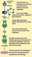
Mast cells contain metachromatic granules that store inflammatory mediators, including rich in histamine and heparin. Mast cells react to allergens and other degranulating agents with sequential exocytosis (description). Mast cells function in wound healing, in autoimmune disorders, and in the inflammatory and immune responses, including allergic reactions and anaphylaxis. (image at left - click to enlarge).

Mast cells are morphologically similar to basophils and both express CD34, though they may have different bone marrow precursors. Unlike basophils, masts cell circulate in an immature form, maturing only in tissue sites where they are resident. Mast cells are found in connective tissue of most organs, and in mucosal tissue.

Mast cells can be activated when an allergen binds to the IgE that is already coating the cell. Allergens are typically proteins or polysaccharides that bind to the Fab segment of the cognate IgE coating the mast cell surface. Crosslinking of two or more IgE molecules is required to activate the mast cell when steric changes disturb the cell membrane structure, triggering an intracellular cascade that activates the mast cell. Although this reaction is most often encountered in allergic reactions, it apparently evolved as a defense system against intestinal parasitism, such as tapeworm infestations.

Mast cells express pattern-recognition receptors plus high-affinity receptor (FcεRI) for IgE, which ligates specific IgE molecules irreversibly.
On activation, mast cells degranulate, releasing preformed mediators into the interstitium:
● histamine
● proteoglycans, mainly heparin (anticoagulant)
serine proteases
● freshly synthesized lipid mediators (eicosanoids)  Eicosanoid Actions :
_prostaglandin D2 (PGD2)
_ ● leukotriene C4 (LTC4)
cytokines

 Complement Receptors  Cytokines  Eicosanoid Actions  Fc receptors  Immune Cytokines  Immunoglobulins  Interferons  Scavenger Receptors  Toll-like Receptors

Top

tags

Labels: , , , , , , , ,

| 0 Guide-Glossary

leukocytes

Leukocytes are white blood cells (WBCs) – left b=basophil, e=eosinophil, m=monocyte, n=neutrophil.

Leukocytes are subdivided according to presense or lack of granulation:

granulocytes generated by granulopoiesis :
--- basophilsmast cells
--- eosinophils
--- neutrophils

agranulocytes
-lymphocytes generated by lymphopoiesis
---B lymphocytesplasma cells
---T lymphocyteshelper T cells, killer T cells
--- ○ natural killer cells → lymphoid dendritic cells

- ○ agranulocytes generated by monocytopoiesis
---monocytesmacrophages
----------------.myeloid dendritic cells

Tables  Fc receptors  Immune Cytokines  Immunoglobulins

æ Archaea & Eubacteria ¤ Cancer Cell Biology ~ Chemistry of Life  Diagrams & TablesEnzymesEvo Devo Molecular Biology φ Molecules ō Organics ››› Pathways ▫▫ Virus

Labels: , , , , , , , , ,

| 0 Guide-Glossary

lymphoid system

Components of the lymphoid system are:
● immune cells – B cell lymphocytes and plasma cells, dendritic cells, granulocytes, macrophages, monocytes of mononuclear phagocyte system (MPS), T cell lymphocytes
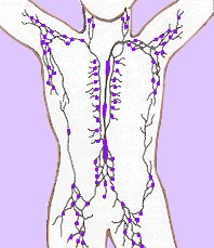
● lymph ducts and vessels and lymph nodes (right)
[] histopathology []
● lymphoid organs including reticuloendothelial system – bone marrow, (lymph nodes), mucosa-associated lymphoid tissue, Peyer's patches, spleen, thymus, tonsils, vermiform appendix

The reticuloendothelial system or mononuclear phagocytic system comprises a range of cells that are capable of phagocytosis, including macrophages and monocytes. Phagocytosis is an innate immune process, and is not an adaptive immune process. The phagocytic cells either circulate in the blood or are attached to various connective tissues such as pulmonary alveoli, liver sinusoids, skin, spleen, and joints.

The RES functions to provide phagocytic cells for both the inflammatory response and immune responses (primary RES) and to remove pathogens and senescent cells from circulation (secondary RES)

The reticuloendothelial system (RES) includes:
● primary (central) lymphoid production organs – bone marrow, thymus
● secondary (peripheral) lymphoid function organs – circulating monocytes, histiocytes located in many tissues, Kupffer cells of the liver, "Littoral cells" of the spleen, mucosa-associated lymphoid tissue (MALT), which is subdivided into bronchus-associated lymphoid tissue (BALT) and gut-associated lymphoid tissue (GALT), Peyer's patches.

Tables  Fc receptors  Immune Cytokines  Immunoglobulins

o-o index of tissue micrographs [] germinal centers [] fluorescence microscopy dendritic cell uptake of dying cells within spleen [] micrograph red pulp of spleen [] micrograph splenic red pulp [] micrograph spleen cells (mouse) DAPK2 stained [] micrograph white pulp, splenic nodule [] micrograph white pulp infiltrate with Langhans giant cell [] histopathology spleen Gaucher's disease [] micrograph gallery cell surface antigens [] Virtual Histology Main []


Top

Labels: , , , , , , , ,

| 0 Guide-Glossary

monocytes

Monocytes are considered to be immature macrophages, and the two types have been considered part of the reticulo-endothelial system (RES) or mononuclear phagocyte system (MPS).

Monocytes play a central role in coordinating immune responses by secreting cytokines and prostaglandins. Cytokines, particularly IL-1, amplify the antigen-induced activation of T cells, whereas released prostaglandins such as PGE2 are potent inhibitors of activation.

Monocytopoiesis takes place in the bone marrow:
stem cell → committed stem cell (common myeloid progenitor) → monoblast → promonocyte → monocyte (bone marrow) → monocyte (peripheral blood) → macrophage or myeloid dendritic cell (tissue).

Monocyte differentiation in the bone marrow requires 1.5 to 3 days. Three glycoprotein growth factors initiate the bone marrow differentiation of macrophages from uni- and bipotential progenitor cells. IL-3 controls the progression from pluripotential stem cell to myeloid-restricted progenitor. IL-3 generates differentiated progeny of all myeloid lineages, and as IL-3-responsive progenitors differentiate, they became responsive to GM-CSF and M-CSF, the two growth factors that determine monocyte/macrophage-restricted progeny. Following commitment to their lineage, monocytes and macrophages remain dependent on these growth factors for continued proliferation and viability. TNF-α, a member of the TNF-receptor superfamily, has also been implicated in growth regulation for macrophage precursors.

Some neutrophilic granules form in the monocyte cytoplasm during development, but these are fewer than those of neutrophil (neutrophilic granulocytes). Circulating monocytes possess migratory, chemotactic, pinocytic, and phagocytic capabilities, in addition to having receptors for IgG Fc-domains (FcR) and iC3b complement. Following migration into tissues, monocytes undergo further differentiation to become multifunctional tissue macrophages.

Tables  Fc receptors  Immune Cytokines  Immunoglobulins

æ Archaea & Eubacteria ¤ Cancer Cell Biology ~ Chemistry of Life  Diagrams & TablesEnzymesEvo Devo Molecular Biology φ Molecules ō Organics ››› Pathways ▫▫ Virus

Labels: , , , , ,

| 0 Guide-Glossary

. . . since 10/06/06
Google