Macrophages (and
dendritic cells) are distributed in peripheral tissues where they eliminate invading foreign substances and are therefore responsible for
innate immunity.
▼:
acquired immunity :
alveolar macrophages :
APC :
exudate macrophages :
free and fixed macrophages :
histiocytes :
IL-1 :
inflammatory macrophages :
Kupffer's cells :
Langerhans's cells :
leukocyte immigration :
life-span :
mononuclear phagocyte system :
MPS :
normal macrophages :
peritoneal, pleural macrophages :
phagocytes :
RES :
reticulo-endothelial system :
tissue renewal :▼
Macrophages are mature, tissue-differentiated
monocytes of the
reticulo-endothelial system (RES) or
mononuclear phagocyte system (MPS). Since macrophages are derived exclusively from
monocytes they exhibit similar properties. The term
exudate macrophages designates the developmental stage and not the functional state.
Inflammatory macrophages are found in exudates, where they may be characterized specific markers, such as peroxidase activity.
Normal macrophages include macrophages located in tissues that include:
● connective tissue –histiocytes
● liver sinusoids – Kupffer's cells
● lung – alveolar macrophages
● lymph nodes – free and fixed macrophages
● spleen – free and fixed macrophages
● bone marrow – fixed macrophages
● serous fluids –pleural and peritoneal macrophages
● skin – histiocytes, Langerhans's cell
Macrophages trigger
acquired immunity by capturing foreign (
exogenous)
antigens, which they ingest in cellular
lysosomes. [
im] Post-hydrolysis, the fragmented antigens are displayed on the cell surface together with macrophage proteins (
APC). A number of
C-type lectins are specifically expressed on macrophages and
dendritic cells.
Coated with fragments of foreign
antigens, macrophages
migrate to
secondary lymphoid organs, where they
present the antigens to
T lymphocytes. This process sensitizes the
T cells to recognize antigens.
Macrophages are ubiquitously distributed mononuclear
phagocytes responsible for numerous
homeostatic,
immunological, and
inflammatory processes. Their wide tissue distribution enables them to provide an immediate defence against foreign elements
prior to
leukocyte immigration. Macrophages participate in both specific immunity via
antigen presentation and in IL-1 production and nonspecific immunity against bacterial,
viral, fungal, and
neoplastic pathogens, so macrophages display a range of functional and morphological phenotypes.
The
life-span of macrophages ranges from 6 to 16 days. Under normal, steady-state conditions, tissue macrophages are renewed by local proliferation of
progenitor cells rather than by monocyte influx into tissue, though invagination of monocytes does occur.
▲:
acquired immunity :
alveolar macrophages ф
antibodies ф
antigen :
APC ф
APCs ф
B cells ф
blood ф
dendritic cells :
exudate macrophages :
free and fixed macrophages ф
granulocytes ф
hematopoiesis :
histiocytes :
IL-1 :
inflammatory macrophages ф
inflammatory response ф
immune cytokines ф
immune response :
Kupffer's cells :
Langerhans's cells :
leukocyte immigration ф
leukocytes :
life-span ф
leukocytes ф
lymphocytes ф
lymphokines ф
lymphoid system ф
migration ф
monocytes :
mononuclear phagocyte system :
MPS :
normal macrophages :
peritoneal, pleural macrophages :
phagocytes ф
phagocyte ф
receptors :
RES :
reticulo-endothelial system ф
T cells :
tissue renewal :▲
[] Macrophage in the process of surrounding tumor cell, artist's impression -
cellular_macrophage [] sem
Macrophage []
sem "walking macrophage" []
sem activated macrophage phagocytosing bacteria []
sem alveolar macrophage attacking E. coli []
tem macrophage-eosinophil []
micrograph macrophage surrounded by normal plasma cells []
micrograph macrophage & plasma cells []
micrograph erythroid island central macrophage []
micrograph foamy alveolar macrophage []
immunofluorescence tubulin in macrophage []
micrograph lymph node []
micrograph lymphoid follicle with germinal center H&E []
micrograph normal spleen []
micrograph macrophage & neutrophils in spleen []
thymus micrograph gallery []
macrophage attacking bacterium []
phagocytic embrace []
cartoon macrophage attacks []
Tables
Fc receptors
Immune Cytokines
Immunoglobulinsanimations Џ
Neutrophil Џ
Platelet Response Џ
Atopic Dermatitis Џ beautiful Flash 8
animation - inner life of the cell and
Interpretation: Inner Life of the Cell Џ
æ
Archaea & Eubacteria ¤
Cancer סּ
Cell Biology ~
Chemistry of Life
Diagrams & Tables ♦
Enzymes ₪
Evo Devo ○
Molecular Biology φ
Molecules ō
Organics ›››
Pathways ▫▫
Virus ▲
Top ▲
Labels: dendritic cells, inflammation, macrophage, monocyte, mononuclear, phagocytes, reticuloendothelial
|


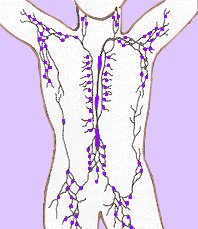
 Components of the lymphoid system are:


