Immunology

basophils

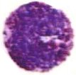
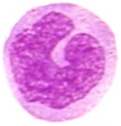
Basophils are granulocytes packed with granules that stain basic (purple with H&E). The nucleus is bilobed, and the metachromatic granules contain sulfated glycosaminoglycans as well as vasoactive compounds – histamine and proteoglycans. Lysosomal arylsulfatase is found in granules of developing basophils in bone marrow. Basophil granules are surrounded by a unit membrane and contain particles which are uniform in size across the same granule yet vary in size in different granules within the same cell. Some granules reveal a homogeneous texture and/or "myelin" figures. [s] Though basophils and mast cells share morphological features, the appearance of most basophil granules differs from the ultrastructure of human mast cell granules.

Activated basophils release proinflammatory histamine and proteoglycans from granules, and synthesize then secrete leukotrienes and cytokines (particularly IL-4). Histamine and IL-4, which is associated with production of IgE, are involved in allergic reactions.

Basophils are the least common granulocyte, representing about 0.5% to 1% of circulating leukocytes. A low basophil count combined with a low neutrophil count almost always portends leukemia.

Tables  Fc receptors  Immune Cytokines  Immunoglobulins .

tags

Labels: , , , , , , , , , , , , ,

| 0 Guide-Glossary

dendritic cells

Dendritic cells and their immature counterparts, Langerhans cells (LC), are highly specialized, professional antigen-presenting cells (APC) located in the skin, mucosa, and lymphoid tissues.

adhesion : APC activities : clonal expansion B cells : cytokines : DC types : disorders : ectopic FDC-formation : follicular dendritic cells (FDC) : FDC networks : generating germinal centers : germinal centers : immature dendritic cells : immune regulators : immunological synapse : interferon producing cells : lymphoid dendritic cells : maturation : morphology : myeloid dendritic cells : pDC : plasmacytoid dendritic cells (PDC, IPC) : precursor dendritic cells : regulators : Th1 and Th2 stimulation : TLRs : types of DC : veiled cells

Immune dendritic cells are named for their morphology (long surface projections), and bear no relationship to neurons. Immature dendritic cells are also called 'veiled cells' because they display large cytoplasmic 'veils' rather than dendrites. DC and LC play a key role in the induction phase of contact allergenicity.

As key regulators of immune responses, dendritic cells (DC) stimulate lymphocytes to perform cell-mediated and humoral immune responses against pathogens and tumor cells. DCs can also educate T cells to tolerate self-antigens, thereby minimizing autoimmune reactions.

Types of dendritic cell
follicular dendritic cells (origin?) – FDC
lymphoid dendritic cells (lymphopoiesis) –
myeloid dendritic cells (monocytopoiesis) – MDC1, MDC2
plasmacytoid dendritic cells – PDC, IPC – the major producers of type I interferon (IFN)

Immature, precursor dendritic cells (pDC) circulate throughout the body, migrating to lymphocyte rich tissues (such as spleen and lymph nodes) upon stimulating encounter with antigen. The dendritic cells internalize the antigen, then digest, and externalize the fragmented antigen that they present to lymphocytes in MHC-peptide complexes, expressing markers that stimulate lymphocyte activation. Dendritic cells are the most effective antigen presenting cells. Follicular dendritic cells stimulate differentiation of B cells, monocytopoietic lineages (pDC1) stimulate differentiation of Th1 cells, lymphopoietic dendritic cells (pDC2) induce differentiation of Th2 cells. Plasmacytoid cells produce type 1 interferon (IFN-α, β, Ω) and can mature into dendritic cells that link innate and adaptive immune responses.

A variety of factors operate in antigen recognition and processing by immature (precursor) dendritic cells and in the maturation of immature cells. Toll-like receptors on the surfaces of precurson dendritic cells recognize microbial components and induce the differentiation of dendritic cell precursors. GM-CSF and IL-4 stimulate the maturation of monocytopoietic pDC1, while IL-3 stimulates the differentiation of pDC2. The transition to mature dendritic cells down-regulates those factors that were involved in antigen internalization, while up-regulating the expression of MHC, costimulatory molecules that participate in lymphocyte activation, adhesion molecules, and specific cytokines and chemokines.

Adhesion molecules enhance direct interactions between T cells and dendritic cells (immunological synapse). Dendritic cell stimulation of formation of Th1 and Th2 cells appears to be regulated by negative feedback. Th1 production of interferon-γ blocks the further stimulation of Th1 differentiation by DC1 cells. Th2 production of IL-4 kills the dendritic cell precursors that contribute to Th2 cell creation. Thus, although IL-4 stimulates Th2 differentiation, the promotion of Th2 cell formation by DC2 cells does not appear to involve IL-4. Costimulatory receptors CD80 and CD86 expressed by mature dendritic cells activate T cells in concert with the recognition of antigen/MHC by the T cell receptor. The secretion of IL-12 by dendritic cells stimulates T cell responses, in particular the differentiation of Th1 cells, which produce interferon-γ and other inflammatory cytokines.

Follicular dendritic cells are stromal cells unique to primary and secondary lymphoid follicles. FDCs express all three types of complement receptors as well as Ig-Fc receptors, through which antigen-antibody immune complexes are retained. FDCs present native antigens to potential memory B cells, of which only those coated with high affinity B cell receptors (BCR) are able to bind.

Recirculating resting B cells migrate through the FDC networks. Antigen-activated B cells undergo clonal expansion within the FDC networks in a T cell-dependent fashion, generating germinal centers. Evidence suggests the presence of two types of dendritic cells within human germinal centers: (i) the classic FDCs that express DRC-1, KiM4, and 7D6 antigens represent stromal cells; and (ii) the newly identified CD3-CD4-CD11c- germinal center dendritic cells (GCDC) represent hematopoietic cells that may be analogous to antigen-transporting cells of mice.

Within germinal centers, B cells undergo somatic hypermutation, positive and negative clonal selection, isotype switching and differentiation into high-affinity plasma cells and memory B cells. Adhesion between FDCs and B cells is mediated by ICAM-1 (CD54)-LFA-1(CD11a) and VCAM-VLA-4. T cells may interact with FDCs in a CD40/CD40-ligand-dependent fashion.

Ectopic FDC-formation is found in a number of autoimmune diseases and/or chronic inflammatory situations, suggesting that FDC development is not restricted to secondary lymphoid organs, but rather that local conditions drives a precursor cell type into FDC-maturation. The precursor of FDCs has presently not been identified, but data suggests a close relation to fibroblast-like cells. [s] It was initially believed that all DCs were of myeloid origin until several recent studies demonstrated that some DCs could also be efficiently generated from lymphoid-restricted precursors. FDCs appear to be involved in the growth of follicular lymphomas and in the pathogenesis of HIV infection.[pm]

Lymphoid dendritic cells are of lymphopoietic origin, and IL-3 stimulates the differentiation of pDC2 cells into DC2 cells, which stimulates differentiation of Th2 cells, which secrete the lymphokine interleukins 4, 5, 10, and 13. (IL-4, IL-5, IL-10, IL-13)

Myeloid dendritic cells are of monocytopoietic origin, and the maturation of precursor cells (pDC1) is stimulated by GM-CSF, and IL-4. Mature DC1 cells secrete interleukin 12 (IL-12), which acts through the JAK-STAT pathway to induce Th1 cells to secrete TNF-β (lymphotoxin) and IFN-γ. MDC-1 is the more common subtype, and is a major stimulator of Th1 cell differentiation. MDC-2 is rare, and may function in response to wound infection.

Plasmacytoid dendritic cells (pDC=IPC) are the major producers of type I interferon (IFN) and exhibit the unique ability to link innate and adaptive immune responses, by differentiating into DC capable of stimulating naive T cells and modulating the adaptive immune response. Human plasmacytoid DCs (PDCs) can induce either Th1- or Th2-type immune responses upon exposure to viruses or IL-3, respectively.

Plasmacytoid dendritic cell precursors (pDC) are type 1 interferon-(α, β, Ω)-producing cells (IPCs) that comprise 0.2%-0.8% of peripheral blood mononuclear cells (humans, mice). IPCs display plasma cell morphology, selectively express Toll-like receptor (TLR)-7 and TLR9, and rapidly secrete massive amounts of type 1 interferon following viral stimulation. IPCs promote the function of natural killer cells, B cells, T cells, and myeloid DCs through type 1 interferons (IFN) during an antiviral immune response. Later in viral infection, IPCs differentiate into a unique type of mature dendritic cell, which directly regulates the function of T cells and thus links innate and adaptive immune responses. [s, ] [fft]

images [] sem dendritic cell and T cell [] micrograph Langerhans cells [] PKC bII signaling in dendritic cells [] micrograph gallery dendritic cells [] photomicrograph dendritic cells interacting with yeast (lilac) [] photomicrograph Human Dendritic cell (labelled with anti MHC class-I FITC) presenting Influenza antigens to T-lymphocytes.
videos Џ Nature video library index Q Џ

ф activation ф affinity maturation ф anergy ф antibodies ф antigen ф APCs ф autoimmunity ф B cells ф bloodbone marrow ф CD ф cellular response ф class-switch recombination ф clonal selection ф complement system ф costimulation ф helper T cell ф hematopoiesis ф humoral immunity ф immune cytokines ф immune response ф immune tolerance ф inflammatory response ф interferons ф isotype switching ф killer T cells ф lymphocytes ф lymphokines ф lymphoid system ф lymphopoiesis ф macrophages ф MHC ф monocytopoiesis ф pattern-recognition receptors ф phagocyte ф plasma cells ф receptors ф signaling ф somatic hypermutation ф surface receptors ф T cells ф thymus

Tables  Complement Receptors  Cytokines  Fc receptors  Immune Cytokines  Immunoglobulins  Interferons  Scavenger Receptors  Toll-like Receptors

IPC: professional type 1 interferon-producing cells and plasmacytoid dendritic cell precursors. [Annu Rev Immunol. 2005]
Plasmacytoid dendritic cell precursors/type I interferon-producing cells sense viral infection by Toll-like receptor (TLR) 7 and TLR9. [Springer Semin Immunopathol. 2005] PMID: 15592841 [Free Full Text]
Natural type I interferon-producing cells as a link between innate and adaptive immunity. [Hum Immunol. 2002] PMID: 12480256
Thrombopoietin cooperates with FLT3-ligand in the generation of plasmacytoid dendritic cell precursors from human hematopoietic progenitors. [Blood. 2004] PMID: 14670916
Flexibility of mouse classical and plasmacytoid-derived dendritic cells in directing T helper type 1 and 2 cell development: dependency on antigen dose and differential toll-like receptor ligation. [J Exp Med. 2003] PMID: 12515817
Roles of toll-like receptors in natural interferon-producing cells as sensors in immune surveillance. [Hum Immunol. 2002]

tags

Labels: , , , , , , , , , , , ,

| 0 Guide-Glossary

immune cytokines

Cytokines are small proteins that regulate and mediate immunity, inflammation, and hematopoiesis. They are secreted de novo in response to immune stimuli, and usually act briefly, locally, at very low concentrations (the exception being endocrine action at distant cells). All cytokines possess four conserved cysteine residues, and are divided into two families based upon the contiguity (CC) or separation of cysteine residues (CXC).

The orchestration and regulation of cytokine production during inflammatory responses constitutes a key determinant of both the resolution of challenge and the limitation of host tissue damage. Cytokines bind to specific membrane receptors, which then signal the cell via second messengers, often tyrosine kinases, to alter cellular activity (gene expression). Cytokines are often produced in cascades.

Tables  Immune Cytokines  Fc receptors  Immunoglobulins  Interferons

actions : chemokine : chemokine family receptors : classification of cytokines : colony-stimulating factors (CSF) : cytokine cascade : cytokine effects : cytokine receptors : cytokine secreting cells : distance of cytokine action : granulocyte colony-stimulating factor (G-CSF) : granulocyte-macrophage colony-stimulating factor (GM-CSF) : hematopoietin family receptors : interleukins, interleukins as largest group : interferon family receptors (IFNR) : largest group of cytokines : lymphokine : lymphotoxins : macrophage colony-stimulating factor (M-CSF) : mechanism of cytokine action : monokine : receptors : search for novel cytokines : target cells : tumor necrosis family receptors
(TNFR) ▼

Actions of cytokines include:
a) up- or down-regulation of the expression of membrane proteins (including cytokine receptors),
b) secretion of effector molecules: histamine release; antibody secretion - IgA, IgG1, and IgE synthesis; IL-1 synthesis; cytokine production; MHC Class II; and CAM expression
c) cellular proliferation
d) chemotaxis of neutrophils, monocytes, and T cells
e) cellular differentiation
f) inflammation
g) phagocytosis
h) death of tumor cells
i) elimination of pathogens

Cytokines are classified according to the cells that produce them:
a) Lymphokines - produced by lymphocytes
b) Monokines - produced by monocytes,
c) Chemokines - chemotactic activities
d) Interleukins - manufactured by one leukocyte to act on other leukocytes.

The cells that produce cytokines include B cells, T cells, dendritic cells, NK, Tc, Th, Th1, Th2, endothelial cells, mast cells, plasma cells, progenitor, marrow stroma, thymus stroma, and tumor cells, along with fibroblasts, leukocytes, monocytes, and macrophages.

Cytokine function may be targetted at:
a) The cells that secrete them (autocrine action),
b) Local cells (paracrine action),
c) Distant cells (endocrine action).

Target cells include: B cells, T cells, NK, Tc, Th, Th2, stem, mast cells, plasma cells, progenitor, tumor cells, phagocytes, neutrophils, monocytes, and macrophages.

Macrophage migration inhibitory factor (MIF) is an inflammatory cytokine secreted by several cell types, including mononuclear cells of the immune system and pituitary cells in the brain. Functional promoter polymorphisms in the MIF gene affect the clinical presentation of systemic sclerosis (SSc, scleroderma) and confer susceptibility to systemic lupus erythematosis (SLE). MIF counteracts cortisol-induced inhibition of inflammatory cytokine secretion, and employs the CD74 receptor.

Cytokines bind to specific membrane receptors:
a) Hematopoietin family receptors - dimers or trimers with conserved cysteines in their extracellular domains and a conserved Trp-Ser-X-Trp-Ser sequence.
The two subunits are i) cytokine-specific, and ii) signal transducing.
Cytokine binding promotes dimerization of the alpha and beta subunits, which then associate with cytoplasmic tyrosine kinases to phosphorylate proteins and activate mRNA transcription. Examples - receptors for IL-2 through IL-7 and GM-CSF.

Colony-stimulating factors (CSFs) are glycoprotein molecules that support growth of hematopoietic colonies. Interleukin 3 (IL-3) exhibits broad activity in the proliferation and differentiation of erythroid, megakaryocytic, and myeloid lineage progenitor cells.

Macrophage colony-stimulating factor (M-CSF, CSF1) acts selectively on the macrophage lineage, while granulocyte colony-stimulating factor (G-CSF) acts selectively on cells of the granulocyte lineage.

Granulocyte colony-stimulating factor (G-CSF) and granulocyte-macrophage colony-stimulating factor (GM-CSF) have pleiotropic activating effects on mature leukocytes, which can improve leukocyte function, facilitating eradication of microbial infections. G-CSF activates neutrophils, while GM-CSF activates neutrophils, eosinophils, and monocyte/macrophages.

G-CSF is a 19.6 kDA glycoprotein that exists in two forms, comprising either 174 or 180 amino-acids. It is produced in a number of different tissues and stimulates granulopoietic proliferation and differentiation in the bone marrow. GM-CSF is a 14.6 kDa globular protein of 128 amino acids that contains two intramolecular disulfide bonds and two potential N-linked glycosylation sites. It is produced by endothelial cells, monocytes, fibroblasts and T-lymphocytes. GM-CSF is a hematopoietic growth factor that stimulates the development of macrophages and neutrophils, and inhibits neutrophil migration while enhancing the functional activity of the mature end-cells. It also promotes the proliferation and development of early erythroid megakaryocytic and eosinophilic progenitor cells.

b) Interferon (IFN) family receptors have the conserved cysteine residues but not the Trp-Ser-X-Trp-Ser sequence, and include the receptors for IFNα, IFNb, and IFNγ.

c) Tumor Necrosis Factor family receptors possess four extracellular domains, and include receptors for soluble TNFα and TNFβ as well as membrane-bound CD40 (important for B cell activation and macrophage activation) and Fas (which signals the cell to undergo apoptosis). TNFα is lymphotoxin β (LT), where the lymphotoxins occur either as homotrimers of LT-α (LT-α3) or as heterotrimers of one LT-α subunit plus two LT-β subunits (LT-α,β2).[s]

d) Chemokine family receptors have seven transmembrane helices and interact with G protein. This family includes receptors for IL-8, MIP-1 and RANTES. Chemokine receptors CCR5 and CXCR4 are used by HIV to preferentially enter either macrophages or T cells.

Cytokines are often produced in cascades, as one cytokine stimulates its target cells to produce additional cytokines. Cytokines are redundant in their activity, in that different cytokines can stimulated similar functions. Different cell types may secrete the same cytokine, or for a single cytokine may act on several different cell types (pleiotropy). Cytokines can also act synergistically with two or more cytokines acting together, or antagonistically with cytokines causing opposing activities.

Interleukins comprise the largest group of cytokines, stimulating proliferation and differentiation of immune cells. The group includes Interleukin 1 (IL-1), which activates T cells; IL-2, which stimulates proliferation of antigen-activated T and B cells; IL-4, IL-5, and IL-6, which stimulate proliferation and differentiation of B cells; Interferon gamma (IFNγ), which activates macrophages; and IL-3, IL-7 and Granulocyte Monocyte Colony-Stimulating Factor (GM-CSF), which stimulate hematopoiesis.

Short half lives, low plasma concentrations, pleiotropy, and redundancy combine to make the isolation and characterization of cytokines difficult. Search for novel cytokines is often conducted at the DNA level, identifying genes that are similar to known cytokine genes.

actions : chemokine : chemokine family receptors : classification of cytokines : colony-stimulating factors (CSF) : cytokine cascade : cytokine effects : cytokine receptors : cytokine secreting cells : distance of cytokine action : granulocyte colony-stimulating factor (G-CSF) : granulocyte-macrophage colony-stimulating factor (GM-CSF) : hematopoietin family receptors : interleukins, interleukins as largest group : interferon family receptors (IFNR) : largest group of cytokines : lymphokine : lymphotoxins : macrophage colony-stimulating factor (M-CSF) : mechanism of cytokine action : monokine : receptors : search for novel cytokines : target cells : tumor necrosis family receptors (TNFR) ▲

Tables  Immune Cytokines  Cell signaling  Receptor Tyrosine Kinases(RTK)  Second Messengers  Phosphate-handling Enzymes  Cell Adhesion Molecules (CAM) .

· adenylyl (adenylate) cyclase ф antibodies ф antigen ф B cells · cadherins · calcium ions · cAMP-dependent protein kinase · CDKs ф cellular response · chemotaxis ф class-switch recombination ф clonal selection ф complement system ф costimulation · cyclin-dependent kinases · cytokine receptors · DAG ф dendritic cells · diacylglycerol · DNA ligases · ERKs · GPCRs · GPCR families ф granulocytes · guanylate cyclases · guanyl cyclase ф helper T cell ф hematopoiesis ф humoral immunity · Ig superfamily ф immune response ф inflammatory response · inositol triphosphate · integrins · IP3 ф leukocytes ф leukocyte adhesion cascade ф lymphokines ф macrophages · MAP kinases ф macrophages ф MHC ф migration · mitogen activated protein kinases ф monocytes ф neutrophils ф pathogens ф pattern-recognition receptors ф phagocyte · phosphatases · phosphodiesterases ф plasma cells · phospolipases · phosphorylation · PKA · PKC · phospholipase C-gamma · protein kinase A · protein kinase C · protein tyrosine kinases (PTKs) ф receptors · receptor tyrosine kinases · second messengers · second messenger cAMP · second messenger cGMP · selectins ф signaling · signal transduction ф surface receptors ф T cells · TNFs · two-component systems ·

Tables  Fc receptors  Immune Cytokines  Immunoglobulins  Interferons  Cell Adhesion Molecules  Cell signaling  Receptor Tyrosine Kinases (RTKs)  Receptor Signal Transduction  Second Messengers 

Labels: , , , , ,

| 0 Guide-Glossary

inflammatory response

Inflammation is a signal-mediated response to cellular insult by infectious agents, toxins, and physical stresses. While acute inflammation is important to the immune response, chronic inappropriate inflammation can cause tissue destruction (autoimmunity, neurodegenerative, cardiovascular disease).

acute phase : agents : cytokines : events : pro-inflammatory cytokines : sequence : signs/symptoms

Symptoms & Signs: Inflammation is variably accompanied by fever (pyrogenesis), redness (rubor), swelling (turgor), pain (dolor), and tissue/organ dysfunction (functio laesa).

The sequence of inflammatory events is:
● insult by trauma or pathogenacute phase reaction
_ ● platelet adhesion, vasoconstriction of efferent vessels
_cytokine induced afferent vascular dilatation (vasodilation causing increased blood flow (redness, local heat) to infected/damaged area
_ ● activation of complement system, clotting system, fibrinolytic system, and kinin system
__leukocyte adhesion cascade
__ ● endothelial gaps increase vascular permeability and allow extravasation of serum proteins (exudate) and leukocytes (→ neutrophilsmacrophageslymphocytes) with resultant tissue swelling
___phagocytosis of foreign material with pus formation

The inflammatory response is part of the innate immune response, and employs cellular and plasma-derived agents (pathway):
complement system
interferons (IFN)
cytokines, lymphokines, monokines
prostaglandins and leukotrienes – arachidonic acid derivatives
● platelet activating factor (PAF)
● histamine
kinins (bradykininpain)

Pain-evoking mediators include proinflammatory cytokines, chemokines, protons, nerve growth factor, and prostaglandins, which are produced by invading leukocytes or by local cells.

Acute phase proteins fluctuate in response to tissue injury and infections. They are synthesized (by hepatocytes) in response to pro-inflammatory cytokines and include:
C-reactive protein (CRP),
mannose-binding protein,
complement factors,
alpha-1 acid glycoprotein,
alpha 1-antitrypsin,
alpha 1-antichymotrypsin,
alpha 2-macroglobulin,
serum amyloid P component (SAP, amyloid),
haptoglobins (alpha-2-globulins),
ceruloplasmin,
complement components C3, C4 ,
coagulation factors (fibrinogen, prothrombin, factor VIII, von Willebrand factor, plasminogen)● ferritin

Pro-inflammatory cytokines include IL-1, IL-6, IL-8, TNF-α (tumor necrosis factor alpha), and TNF-β (lymphotoxin α, LT).

In response to infection, macrophages secrete IL-1 and TNFs, which are broad-spectrum cytokines that stimulate inflammatory responses of neutrophils, fibroblasts, and endothelial cells. The fibroblasts and endothelial cells respond to IL-1 and TNF by recruiting more immune cells to the site of inflammation.

Pain:
When tissue is destroyed or invaded by leukocytes in inflammation, numerous mediators are delivered by the circulation and/or liberated from resident and immigrated cells at the site. Proalgesic mediators include proinflammatory cytokines, chemokines, protons, nerve growth factor, and prostaglandins, which are produced by invading leukocytes or by resident cells. Less well known is that analgesic mediators, which counteract pain, are also produced in inflamed tissues. These include anti-inflammatory cytokines and opioid peptides. Interactions between leukocyte-derived opioid peptides and opioid receptors can lead to potent, clinically relevant inhibition of pain (analgesia). Opioid receptors are present on peripheral endings of sensory neurons. Opioid peptides are synthesized in circulating leukocytes, which migrate to inflamed tissues directed by chemokines and adhesion molecules. Under stressful conditions or in response to releasing agents (e.g., corticotropin-releasing factor, cytokines, noradrenaline), leukocytes can secrete opioids. They activate peripheral opioid receptors and produce analgesia by inhibiting the excitability of sensory nerves and/or the release of excitatory neuropeptides. This review presents discoveries that led to the concepts of pain generation by mediators secreted from leukocytes and of analgesia by immune-derived opioids.
Leukocytes in the regulation of pain and analgesia. Rittner HL, Machelska H, Stein C. J Leukoc Biol. 2005 Dec;78(6):1215-22. Epub 2005 Oct 4. [Free Full Text Article]

[] inflammatory initiation - skin Џ animation of leukocyte adhesion Џ

Tables  Fc receptors  Immune Cytokines  Immunoglobulins  Cell Adhesion Molecules  Cell signaling  Receptor Tyrosine Kinases (RTKs)  Receptor Signal Transduction  Second Messengers 

▲ф agents : cytokines : events : pro-inflammatory cytokines : sequence : signs/symptoms ▲ф

Top

tags

Labels: , , , , , , , , ,

| 0 Guide-Glossary

interferons

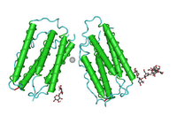
Interferons are immune cytokines that are classified as type I, II, or III according to the receptors through which they signal. Interferon (INF) family receptors have conserved cysteine residues and include the receptors for IFN-α, IFN-β, and IFN-γ.

● IFN type I – bind to the IFN-α receptor (IFNAR) that consists of IFNAR1 and IFNAR2 chains – subtypes of IFN-α, IFN-β (image at left) , IFN-κ, IFN-δ, IFN-ε, IFN-τ, IFN-ω, IFN-ζ (limitin)

● IFN type IIIFN-γ receptor (IFNGR) complex comprising two subunits each of molecules designated IFNGR1 and IFNGR2 – singel type IFN-γ (image at right)

● IFN type III –receptor complex comprising IL10R2 (CRF2-4) plus IFNLR1 (CRF2-12) – types IFN-λ1(IL29), IFN-λ2 (IL28A), IFN-λ3 (IL28B) (image)

Table  Interferons

Cellular secretion of interferon is stimulated by:
● constituents such as CpG DNA from microbial pathogensbacteria (endotoxin, flagellin, CpG) and viruses (viral glycoproteins, viral RNA, CpG)
mitogens
● other cytokines, such as the interleukins (1, 2, 12), TNFs, and colony-stimulating factors

Interferon actions:
● anti-tumor
● antiviral
● activation of macrophages and NK cells
presentation of microbial peptides to T cells through enhancement of MHC classes I and II

 Fc receptors  Immune Cytokines  Immunoglobulins  Interferons

Labels: , , ,

| 0 Guide-Glossary

lymphokines

Lymphokines comprise that subset of cytokines produced by T lymphocytes, whereas monokines are secreted by monocytes. Modern usage employs the more general term 'cytokine'.

Lymphokines include:
colony-stimulating factors (CSFs), including GM-CSF
interferons (IFNs) - IFNγ
interleukins IL-1 to IL-8, IL-10, IL-13
● macrophage inflammatory protein-1 beta (MIP-1β)
● neuroleukin (lymphokine product of lectin-stimulated T cells)[s]
● osteoclast-activating factor
● platelet-derived growth factor (PDGF)
● transforming growth factor beta (TGFβ)
tumour necrosis factor-alpha (cachectin) (TNFα)
tumour necrosis factor-beta (TNFβ, lymphotoxin α, LT)

Actions of lymphokines include
activates B cells, inhibits macrophage function – IL-10
activation of neutrophils, eosinophils, and monocyte/macrophagesGM-CSF
● bone resorption – osteoclast activating factor
bone marrowgrowth and differentiation of immune cells – IL-3
B cell growth and differentiationIL-4
B cell differentiation, activates some microphages (PMN) – IL-5
costimulator of T cells, induces growth in B cellsIL-6
inflammation, fever, catabolism and cachexia, activation of some microphages – TNFs
hematopoiesis stimulators – IL-3, IL-7, GM-CSF
macrophage-activating activity (MAF) – IFN-γ
● stimulates proliferation of activated T and B cells – IL-2
● inhibits T cell growth, activates macrophagesTGFβ

tags

Labels: , , , , , ,

| 0 Guide-Glossary

monocytes

Monocytes are considered to be immature macrophages, and the two types have been considered part of the reticulo-endothelial system (RES) or mononuclear phagocyte system (MPS).

Monocytes play a central role in coordinating immune responses by secreting cytokines and prostaglandins. Cytokines, particularly IL-1, amplify the antigen-induced activation of T cells, whereas released prostaglandins such as PGE2 are potent inhibitors of activation.

Monocytopoiesis takes place in the bone marrow:
stem cell → committed stem cell (common myeloid progenitor) → monoblast → promonocyte → monocyte (bone marrow) → monocyte (peripheral blood) → macrophage or myeloid dendritic cell (tissue).

Monocyte differentiation in the bone marrow requires 1.5 to 3 days. Three glycoprotein growth factors initiate the bone marrow differentiation of macrophages from uni- and bipotential progenitor cells. IL-3 controls the progression from pluripotential stem cell to myeloid-restricted progenitor. IL-3 generates differentiated progeny of all myeloid lineages, and as IL-3-responsive progenitors differentiate, they became responsive to GM-CSF and M-CSF, the two growth factors that determine monocyte/macrophage-restricted progeny. Following commitment to their lineage, monocytes and macrophages remain dependent on these growth factors for continued proliferation and viability. TNF-α, a member of the TNF-receptor superfamily, has also been implicated in growth regulation for macrophage precursors.

Some neutrophilic granules form in the monocyte cytoplasm during development, but these are fewer than those of neutrophil (neutrophilic granulocytes). Circulating monocytes possess migratory, chemotactic, pinocytic, and phagocytic capabilities, in addition to having receptors for IgG Fc-domains (FcR) and iC3b complement. Following migration into tissues, monocytes undergo further differentiation to become multifunctional tissue macrophages.

Tables  Fc receptors  Immune Cytokines  Immunoglobulins

æ Archaea & Eubacteria ¤ Cancer Cell Biology ~ Chemistry of Life  Diagrams & TablesEnzymesEvo Devo Molecular Biology φ Molecules ō Organics ››› Pathways ▫▫ Virus

Labels: , , , , ,

| 0 Guide-Glossary

. . . since 10/06/06
Google