Immunology

helper T cell

Helper T cells, T helper cells (Th), effector T cells circulate throughout the body where they interface with MHC class II protein on other cells, determining whether the MHC class II is presenting 'self' or 'non-self protein' (antigen). MHC class II receptors are located on the surface of professional antigen presenting cells (APCs), which display epitope proteins – exogenous antigen or fragmented angtigen from phagocytosed cells – on their surfaces.

When a helper T cell is activated by contact with antigen, it enters the cell cycle in addition to producing lymphokines and chemokines. Th cells direct antibody class switching in B lymphocytes, orchestrate activation and growth of cytotoxic T cells, and maximize the bactericidal activity of phagocytes (macrophages).

Naïve B lymphocytes each have one of millions of distinct surface antigen-specific surface receptors, yet have not encountered their specific, cognate antigen. With a life-span of only a few days, many B cells die without ever encountering their cognate antigen. Naïve B cells are stimulated when the BCR binds to its cognate antigen. This antigen-Ig binding must be coupled with a signal from a helper T cell in order to activate the B cell.

Helper T cells mostly carry the CD4 surface protein, though a few carry CD8. The CD4 receptor triggers targetting by HIV, which determines the crippling effect of HIV on the immune system.
Subsets of Th cells are defined by the class of cytokine that they secrete upon activation:
Th1 – produce copious amounts of IL-2 and IFN-γ.
Th2 – particularly effective at stimulating B cells through secretion of IL-4, IL-5, and IL-6.
Th3 – produce cytokine transforming growth factor-beta (TGF-β) and IL-10.


Th1 cells are the more effective antiviral agents by virtue of their secretion of interferons (IFN-γ).  Immune Cytokines  Interferons

The cytokines produced by the two Th subsets perform cross-regulatory role. An activated Th2 cell secreting IL-4,-5,and 6 downregulates local Th1 cells in the neighborhood, whereas Th1 cytokines downregulate Th2 responses.

Tables  Fc receptors  Immune Cytokines  Immunoglobulins

[] diagram - helper T cells and phagocytic response to tumor cells [] diagram - HIV binding via CD4 receptors [] micrograph germinal center with helper T cells [] tem - helper T & B cell []

Labels: , , , , , ,

| 0 Guide-Glossary

isotype switching

Isotype switching is a characteristic feature of the humoral immune response, in which a switch from IgM to other Ig isotypes follows first exposure to an antigen. Affinity maturation ensures that repeated exposures to the same antigen will provoke greater antibody ligating affinity of the antibody secreted by successive generations of plasma cells.

Isotype switching is regulated by T cell-produced immune cytokines, such as interleukin-4 (IL-4), interferon-γ (IFN-γ), and TGF-β, which direct B cells to switch to specific Ig classes.

Tables  Fc receptors  Immune Cytokines  Immunoglobulins

Labels: , , , , , ,

| 0 Guide-Glossary

MHC

The major histocompatibility complex (MHC) encodes molecules displayed on cell surfaces, where they participate in lymphocyte recognition and antigen presentation.

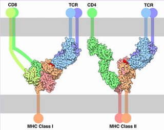
Left - click to enlarge - the presented antigen of the MHC-peptide complex is indicated by red. (Image source Molecule of Month: Illustration by David S. Goodsell of The Scripps Research Institute. Original work of the US Federal Government - public domain.)

The TCR and MHC-peptide complex make primary contact, then CD molecules bind to other portions of the MHC.

Almost ubiquitous, MHC class I interacts with TCRs and CD8 on cytotoxic T cells (CTCs, 'suppressor' T cells, regulator T cells). MHC class II is expressed primarily on cells that interact with pathogenic peptides. MHC II interacts with TCRs and with CD4 on helper T cells and cells that stimulate the immune system.

Proteins encoded by MHC genes identify the molecule as non-self or self. Non-self antigens – partly digested by lysosomes of leukocytes (monocytes or neutrophils) or displayed intact – are carried on the APC surface by class II histocompatibility molecules. Proteins of tumor cells or foreign invaders such as bacteria and viruses are carried by class I histocompatibility molecules. Non-self antigens provoke the immune response.

● Class I – encoded in BCA region – occur on almost every nucleated cell of the body, and are heterodimeric peptide binding proteins, antigen processing molecules such as TAP and Tapasin
● Class II – encoded in D region – occur only on specialized cell types, and are heterodimeric peptide binding proteins, proteins such as MHC II DM, MHC II DQ, and MHC II DP that that modulate peptide loading in the lysosomal compartment
● Class III – encoded between BCA and D regions – complement components such as C2, C4, factor B, and some that encode cytokines (e.g., TNF-α).

The Class I and Class II MHC molecules belong to molecules of the immunoglobulin supergene family, including immunoglobulins, T-cell receptors, CD4, and CD8. The major histocompatibility complex is encoded by several genes located on human chromosome 6 – class I molecules are encoded by the BCA region, while class II molecules are encoded by the D region. Sequences in the region between the BCA and D segments encodes class III molecules, which include some complement components and cytokines.

Human class II molecules are designated HLA-D, for human leukocyte antigen D, and the genes encoding them are also located in the major histocompatibility complex (MHC)

Tables  Fc receptors  Immune Cytokines  Immunoglobulins

æ Archaea & Eubacteria ¤ Cancer Cell Biology ~ Chemistry of Life  Diagrams & TablesEnzymesEvo Devo Molecular Biology φ Molecules ō Organics ››› Pathways ▫▫ Virus

Labels: , , , , ,

| 0 Guide-Glossary

. . . since 10/06/06
Google