Immunology

antibodies

Antibodies are glycoproteins of the immunoglobulin superfamily, and are adhesion-signaling molecules that recognize (bind to) specific antigens. Antibodies are synthesized by B cell-derived plasma cells.

▼: adhesion molecules : antigen binding site : C : CH1-4 : cellular adhesion molecules : complement fixation : complementarity determining regions : constant domains : domains : evolution of immunoglobulins : Fab : Fc : heavy chain : hinge region : Ig supergene family : isotypes : kinase activation : light chain : location of Ig classes : membrane-bound Igs : multimeric structures : tissue location : V : VDJ recombination : VH, VL : variable domains :▼
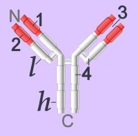
Immunoglobulins (left - click to enlarge) comprise two heavy (h) and two light-chain (l) protein subunits, each of which folds into domains (4 on heavy, 2 on light). These adhesion sites or domains contain one or more folds of 60 to 100 amino acids.

Depending upon the character of the heavy chain, immunoglobulins are divided into five classes – IgG, IgD, IgE, IgA, IgM – that are expressed in different tissues. The classes are further subdivided into isotypes, which have different properties in terms of complement fixation and binding to immunoglobulin (Ig) receptors.

Members of the immunoglobulin supergene family are found as:
● membrane-bound surface receptors of immune-system cells,
cellular adhesion molecules, or
● soluble antibodies (γ-globulins) synthesized by activated B cells.

Membrane-bound Igs have a transmembrane segment and a cytoplasmic C-terminal tail. The 2 β- chains are stabilized into sandwiched β sheets that are adherent by virtue of hydrophobic interactions between disulphide bonds. Igs assume a Y-shaped structure "topped" at the extracellular N-terminals by variable domains (red), with a variable domain at the tip of the heavy chain (1) and the light chain (2), between which lies an antigen binding site (3). The variable regions are coded by pluripotential DNA sequences that can generate thousands of polypeptide sequences capable of adhering to millions of different ligands. Binding is homophilic or heterophilic, including binding to different Igs and to integrins. Both light and heavy chains contain constant domains (white, 4).
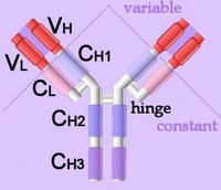
Right - click to enlarge - the heavy chains of IgA, IgD and IgG each have four domains, where those at the N-terminal are variable (VH) and the other three are constant (CH1-3). IgE and IgM have one variable and four constant domains (CH1-4) on the heavy chain. The variable domains are termed Fab, while the constant domains are termed Fc.

The light chains have two domains, one variable domain (VL) at the N-terminal, and one constant (CL) domain.

The antigen binding site lies between VH and VL (shaded lavendar). Most variability is found in three superficial-loop forming regions in the VH and VL domains, which are the complementarity determining regions or CDRs. CDR3 binds antigens and CDR1-2 bind MHCs. CDR3 shows more variation that do either CDR1 or 2.

The domains have related amino acid sequences that possess a common secondary and tertiary structure. This conserved structure is found frequently in proteins involved in cell-cell interactions and is particularly important in immunology. The constant (Fc) regions have complement fixing and Ig receptor binding activity. The hinge region, in IgG, IgA and IgD, is an important sequence of 10-60 amino acids between CH1 and CH2 that confers flexibility on the molecule.

animations Џ B cell selection Џ ELISA test +ve, -ve Џ IgG rotating x- y- axes Џ Rotating mouse IgG2a Molecule (y-axis) Џ somatic recombination of Ig gene Џ spinning IgG1 Kol Џ unfolding (small) IgG . unfolding (large) IgG .

Immunoglobulins attain their enormous variability by splicing components (VDJ recombination) coded in widely scattered sequences of DNA that are located in two different chromosomes. Antigen binding takes place at the heavy chain, which displays enormous variation by virtue of combining 1 of 400 possible variable gene segments with 1 out of 15 diversity segments and 1 out of 4 joining segments. This alternative splicing generates 24,000 possible combinations for the DNA encoding the heavy chain alone. The variable coding segments are assembled together with those for the constant-C segments of the heavy-chain molecule.

Tissue location:
IgA – mucus – gut, respiratory tract
IgD – antigen receptor on B cells
IgE – mast cells – releases histamines in response to allergens
IgG – primary immunity against invading pathogens
IgM – early B cell-mediated response to invading pathogens

Some antibody classes form multimeric structures – pentamers (IgM) and dimers or trimers (IgA). These two isotypes also associate with a small protein called the joining (J) chain required for stabilisation of the complexes.

The immunoglobulin superfamily is evolutionarily ancient, is widely expressed, and is constitutive or long-term up-regulated. Immunoglobulin antibodies are released by activated B cells of the immune system, on which they also act as surface marker proteins. Adherence of immunoglobilins to foreign substances or to cellular invaders may be sufficient to disarm the invader, or the attached antibodies function as attack signal to macrophages and natural killer cells. Adhesion molecules of the immunoglobulin supergene family, activate specific kinases through phosphorylation, resulting in activation of transcription factors, increased cytokine production, increased cell membrane protein expression, production of reactive oxygen species, and cell proliferation.

▲: adhesion molecules ~ adhesion molecules ф antigen : antigen binding site ф APCs ф B cells : C : CH1-4 : cellular adhesion molecules : complement fixation ф complement system : complementarity determining regions : constant domains : domains : evolution of immunoglobulins : Fab : Fc : heavy chain : hinge region ф humoral immunity : Ig supergene family ~ immunoglobulins : isotypes : kinase activationkinases : light chain : location of Ig classes : membrane-bound Igs : multimeric structures ф receptors ф signaling ф surface receptors ф T cells : tissue location ~ tyrosine kinases : V : VDJ recombination ф VDJ recombination : VH, VL : variable domains :▲

Tables  Fc receptors  Immune Cytokines  Immunoglobulins  Cell Adhesion Molecules  Cell signaling  Receptor Tyrosine Kinases (RTKs)  Receptor Signal Transduction  Second Messengers 

Top

tags

Labels: , , , , , , , , , ,

| 0 Guide-Glossary

evolution of immune and coagulation systems

Immune system
The innate immune system is ancient and displays roots roughly one billion years old, deep in the deuterostome branch of the bilaterians (pre-Cambrian). The lectin pathway (MBL - MASP) is homologous to the classical complement pathway, but utilizes opsonin, mannan-binding lectin (MBL, MBP) and ficolins rather than C1q. Diversified ficolins are of particular importance in invertebrates, which lack the adaptive immune response that evolved some 500 million years ago in jawed vertebrates.

Macrophage scavenger receptors appear to mediate important, conserved functions, so it was likely pattern-recognition receptors that arose early in the evolution of host-defense mechanisms. Eicosanoids play a prominent role in inflammatory/immune responses and the evolution of eicosanoid receptors has been analyzed on the basis of amino acid sequences. Eiconasoid receptors are located on a variety of cells, tissues, and organs and can be activated by either non-selective or selective ligands.

The more specific, versatile, memory-capable adaptive immune response evolved more recently, roughly 450 million years ago, and is found in the jawed vertebrates (gnathostomes) but not in invertebrates.

Although the B cells of higher vertebrates lack phagocytic capabilities, it has recently been demonstrated that B cells from teleost (bony) fish and amphibians display potent phagocytic activities. Particle uptake by B cells induced activation of 'downstream' degradative pathways, leading to 'phagolysosome' formation and intracellular killing of ingested microbes. It is most probable that the less-elaborated, restrictive adaptive immune response of fish and amphibians makes the preservation of phagocytosis an evolutionary advantage to B cells in their defence against pathogens. These findings support the idea that B cells evolved from an ancestral phagocytic cell type, providing an evolutionary framework for understanding the close relationship between mammalian B lymphocytes and macrophages.[a, n]

Mast cell degranulation releases histamine and other vasoactive mediators in response to allergens. Although this reaction is most often encountered in allergic reactions, it apparently evolved as a defense system against intestinal parasitism, such as tapeworm infestations.

The versatile immunoglobulin superfamily is evolutionarily ancient, is widely expressed, and is constitutive or long-term up-regulated. Immunoglobulin antibodies are released by activated B cells of the immune system, on which they also act as surface marker proteins. The enormous diversity of antibodies is attributable to the alternative splicing of VDJ recombination.

RAG1 and RAG2, the proteins that mediate VDJ recombination, are closely related to transposases, and it is believed that evolution of the vertebrate genome includes their entry as part of a Transib superfamily transposon.

Blood coagulation employs the same fundamental mechanism in all vertebrates, from the early diverging jawless fishes to mammals.[1]. It has been amply demonstrated that all groups of fish generate thrombin through pathways that:
● utilize vitamin K-dependent factors
● exhibit factor XIII-dependent fibrin cross-linking, and
● manifest a fibrinolysis inhibited by the same antifibrinolytic agents as mammals (13).

(Thrombin-generated fibrin coagulation has not been observed in nonvertebrate chordates or in other invertebrate animals.)

Such a convoluted pathway as the clotting cascade could not have evolved as a single event. Proponents of "intelligent design theory" attempted to monopolize on this fact in order to promote their claims that an intelligent designer (God) must be responsible for the so-called "irreducible complexity" of the coagulation cascade. (Behe is a little more cautious in his wording, but the implied argument is as stated above.) Just as for the claims of irreducible complexity for evolution of the eye and the bacterial flagellum, the argument has been both logically and scientifically refuted.

Scientists realized some time ago that a series of gene duplications must be responsible for the complex set of interactions observed in mammalian clotting. Sequence comparisons of serine proteases led to the suggestion that the contact system of clotting factors ( factors XI and XII, and prekallikrein) must have evolved more recently than some of the other clotting factors and thus would likely be absent in lower vertebrates (4).

The genome sequences (5) for the puffer fish, Fugu rubripes, along with that for the urochordate (sea squirt) Ciona intes (6) have enabled a direct comparison of two early diverging chordates. The genomes confirm that the main lines of the vertebrate clotting pathway were evolved during the less than a hundred million years between the last common ancestor of these two creatures. It is currently believed that 50–100 million years separate the appearances of urochordates (including the sea squirt) and vertebrates. During this interval, the machinery for thrombin-catalyzed fibrin formation was presumably 'concocted by gene duplication and the shuffling about of key modular domains'.[adapted from article]

Talk Origins Evolving Immunity . Evolution of the Immune System, Spring 2005 .

Sequence comparisons of the three homologous polypeptide chains that compose vertebrate fibrinogens (acute phase proteins) imply that the molecule evolved before the divergence of vertebrates and invertebrates. Computer comparisons of various fibrinogen-related sequences indicate that the sea cucumber proteins diverged before the beta-gamma gene duplication.
Presence of a vertebrate fibrinogen-like sequence in an echinoderm. [Proc Natl Acad Sci U S A. 1990]

Coelomocytes increased expression of ferritin mRNA after stimulation. In vertebrates, cytokines can cause changes in iron levels in macrophages. Similarly, echinoderm macrokines produced decreases in iron levels in coelomocyte supernatant fluids. These results suggest that echinoderm ferritin is an acute phase protein and suggest that sequestration of iron is an ancient host defense response in animals.
Evolution of the acute phase response: iron release by echinoderm (Asterias forbesi) coelomocytes, and cloning of an echinoderm ferritin molecule.[Dev Comp Immunol. 2002 Jan;26(1):11-26.]

tags

Labels: , , , , , , , , , , , , , ,

| 0 Guide-Glossary

VDJ recombination

VDJ recombination, also known as antigen receptor gene rearrangement or antigen-independent diversification, is a diversity generating assembly process affecting the variable domain of immunoglobulin and TCR genes.

▼: 12/23 rule : B cells : class-switch recombination : CSR : double strand breaks : E2A encoded proteins : hairpin : HMG-1, HMG-2 : lymphoid-specific components : nonlymphoid-restricted components : RAG1 & RAG2 : RSS : SAGA : SHM : somatic hypermutation : synaptic complex : transposon : VDJ genes :▼

The heavy (H) plus kappa (κ) or lambda (λ) chain combinations of BCRs (H-κ/λ) and the alpha (α) and beta (β) or gamma (γ) and delta (δ) chain combinations of TCRs (αβ or γδ) are encoded by roughly three hundred different gene segments, yet produce an estimated 5 x 10^7 to 10^9 surface receptors (B+T). The segments are scattered on human chromosomes 2, 14, and 22.

Segments:
BCR heavy chains: 51 VH, 27 DH, 6 JH, 9 CH gene segments on human chromosome 14 (93 VDJC). The CH segments are 1 µ (IgM), 1 δ (IgD), 4 γ (IgG), 1 ε (IgE), and 2 α (IgA)
BCR light chains 40 Vκ , 31 Vλ , 5 Jκ , 4 Jλ gene segments on human chromosome 14 (80 VJ)

TCR 50 Vα, 50 Jα, 20 Vβ, 13 Jβ , 2 Dβ gene segments (100 α, 35 β), and a smaller number of γδ gene segments. The κ segments are on human chromosome 2 and the λ segments are on human chromosome 22

Antigen receptor gene rearrangement of variable (V), diversity (D) and joining (J) gene segments generates this enormous repertoire of antigen receptors with different antibody specificities, providing the versatility that is essential to normal immune functioning. It has been estimated that around 10^9 distinct antibody molecules can be generated by VDJ recombination. The genes encoding the variable region domain for the heavy chain lack a complete exon, instead segments encoding the V region are split into arrays of gene segments. Light chain genes are also organised on different chromosomes, but they have no D gene segments. There are 51 functional VH genes and 41 Vk genes. D (diversity) and J (junctional) genes code for amino acids at the carboxyl end of V regions including CDR3.

Each heavy chain is derived from a V, D, J, and C region in a sequence of steps:
1. A D and J sequence are spliced
2. A V segment is spliced to the DJ segment, all intervening Vs and Js are deleted when the random V and J are joined. This brings V, D, and J gene segments together in a translational reading frame at the DNA level, generating a mRNA product: Leader, V, D, J, C, poly A

Each light chain begins with the V and J sequences combined, with a few thousand base pairs separating the J and the C regions. This is then transcribed into a primary transcript, polyadenylated, and the intervening sequence is spliced out. This generates the mRNA product: Leader, V, J, C, poly A.

Antigen-dependent immunoglobulin gene diversification, via somatic hypermutation (SHM), and class-switch recombination (CSR) occur in mature B cells during the humoral immune response. SHM generates point mutations and CSR generates different antibody isotypes by recombination 1, 2.

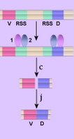
Highly conserved recombination signal sequences (RSS), comprising a heptamer and a nonamer motif with an intervening 12- or 23-bp spacer, enable VDJ recombination of the immunoglobulin and TCR loci involving RSS with different spacers following the 12/23 rule (3). Right - click to enlarge - simplified process of VDJ recombination in which V and D coding segments of immunoglobulin and T cell receptor genes are flanked by short recombination signal sequences (RSS), which are in opposite orientations at the 5' and 3' termini of the coding sequences. That is, RSS are located at the 3' end of each V segment, 3' and 5' ends of each D segment, and at the 3' end of each J segment. The RSS are recognized (1, 2)by a complex of the lymphocyte-specific recombination proteins, recombination activating genes, RAG1 and RAG2 (for recombination activating genes). These enzymes cleave (c) the DNA between the coding sequence and the RSS, creating double-strand breaks (DSB). The broken coding strands are then ligated (j) by nonhomologous end-joining to yield a rearranged gene segment (D-J, then DJ-V):
● a coding join (D-J or V-DJ for heavy chains; V-J for light chains), which is retained, and
● a signal join, formed from a loop of DNA from which has been deleted all the intervening DNA initially present between the 2 gene segments. The signal join segment is discarded.
(For more comprehensive diagrams and legends see here.) Џ AV animations click on thumbnails Џ

RAG1 and RAG2, the proteins that mediate VDJ recombination, are closely related to transposases, and it is believed that evolution of the vertebrate genome includes their entry as part of a Transib superfamily transposon.

Several proteins mediate VDJ recombination (4):
● Lymphoid-specific components of the recombination machinery, RAG-1, RAG-2 together constitute the recombinase. Terminal deoxynucleotidyl transferase (TdT) mediates the incorporation of nontemplate-dependent nucleotides.
● Nonlymphoid-restricted components include DNA-PKcs, Ku70, Ku80, XRCC4, ligase 4, Artemis, and possibly HMG1 and HMG2.

All these proteins are involved in repair of DNA double strand breaks in addition to their rôle in VDJ recombination. The nonlymphoid-specific components probably participate in the processing and joining steps of VDJ recombination. HMG1 and HMG2 are two additional nonlymphoid-specific components that have been implicated in VDJ recombination. Experiments have demonstrated that these proteins increase the in vitro efficiency of cleavage by RAGs (5, 6).

During the initial stages of antigen receptor gene rearrangement, RAG-1/RAG-2 form a complex with the RSS, which is partly stabilized by interactions between the nonamer binding domain of RAG-1 and the nonamer motif. Bridging of 12 and 23 RSS, in a synaptic complex, is critical for DNA cleavage and for it to be facilitated by the DNA bending proteins HMG1 and HMG2. Within the synaptic complex, RAG-1/RAG-2 efficiently introduce a nick at each RSS through a hydrolysis reaction at the heptamer/coding flank border, which generates a 3' hydroxyl end. A transesterification reaction, resembling the mechanism of transpositional recombination, next creates a double strand break as the free 3' hydroxyl of the nicked strand is used in a nucleophilic attack on the opposing strand generating a covalently sealed hairpin intermediate, known as the hairpin coding end (5, 6). [s]

E2A encoded proteins, including E12 and E47, regulate site-specific DNA recombination. The extreme N-terminal domain of E2A has been shown to recruit the co-activator protein complex, SAGA, which contains histone acetylase activity, so it is conceivable that the E2A proteins regulate recombination by promoting locus accessibility. Possible mechanisms by which E2A proteins regulate recombination include localized accessibility, looping, and direct recruitment of RAG proteins.[l]

The VDJ recombination mechanism in jawed vertebrates is catalyzed by the RAG1 and RAG2 proteins, which are believed to have emerged approximately 500 million years ago from transposon-encoded proteins. Although no transposase sequence similar to RAG1 or RAG2 has been found, the approximately 600-amino acid “core” region of RAG1 required for its catalytic activity is significantly similar to the transposase encoded by DNA transposons that belong to the Transib superfamily. It has been demonstrated that recombination signal sequences (RSSs) were derived from terminal inverted repeats of an ancient Transib transposon. Furthermore, the critical DDE catalytic triad of RAG1 is shared with the Transib transposase as part of conserved motifs.[r] These findings refute one of Behe's claims for irreducible complexity of complex biochemical features.

. chromosome 2, chromosome 14, chromosome 22 .

▲:12/23 rule ф antibodies ф antigen »» Basic mechanisms of evolution : B cells : class-switch recombination : CSR : double strand breaks ~ double strand breaks : E2A encoded proteins : hairpin : HMG-1, HMG-2 : lymphoid-specific components : nonlymphoid-restricted components »» point mutation : RAG1 & RAG2 »» Recombination : RSS : SAGA : SHM : somatic hypermutation ~ splicing : synaptic complex : transposon ~ transposons : VDJ genes :▲

Tables  Fc receptors  Immune Cytokines  Immunoglobulins

Top

External : Transposons part 1, transposons part 2 : Barbara McClintock and mobile genetic elements :

Labels: , , , , , , , ,

| 0 Guide-Glossary

. . . since 10/06/06
Google