Immunology

Immunology Overview

Immunology is the study of the immune system, which defends the body against foreign intrusion by pathogens. (right - colorized scanning electron micrograph of red blood cells - erythrocyte, platelet, leukocyte)

The immune system is intimately connected with the hematologic system since white blood cells (leukocytes, including B- and T-lymphocytes) are key players in the lymphoid system. Cellular participants in the immune and inflammatory responses include :
phagocytic cells (dendritic cells, monocytes and macrophages, and granulocytes)
antigen presenting cells (dendritic cells, macrophages, B lymphocytes, helper T cells)
antibody producing cells (plasma cells)
cytotoxic cells (CTL, NK, NKT)
● regulatory cells (APCs, helper T cells, regulatory T cells)
● cells-in-waiting (memory B cells, monocytes, naïve B cells, Tc)
● chemical releasing cells (basophils, eosinophils, neutrophils; mast cells - histamine, cytokines; hepatocytes - complement proteins)

An antigen is any molecule that stimulates an immune response. Most antigens are proteins or polysaccharides, though small molecules coupled to carrier proteins (haptens) can also be antigenic. The segment of an antigenic molecule to which its cognate antibody binds is termed an epitope or antigenic determinant. Immune responses ideally distinguish between self and other. Anergy toward self-targets operates as one self-tolerance mechanism to control the autoreactive cells found in disease-causing autoimmunity.

Immune responses are classifed as passive or active, innate or adaptive, and cellular or humoral.

These categories are not mutually exclusive. For example, both innate and adaptive immune responses employ cellular responses. Similarly, humoral and cellular responses intersect rather than being mutually independent (e.g., helper T cells assist in activation of B cells, opsonization). Unfortunately, some terminology employed in immunology predates understanding of mechanisms, so some commonly used names do not immediately reflect the distinction between cell types (NK cells versus NKT cells) or origins (lymphoid versus myeloid origins of dendritic cells). Similarly revision of chemical terminology has resulted in misleading terminology of biochemical components (e.g., complement C4b•2b was formerly termed C4b•2a).

Passive measures to prevent pathogenic incursions are provided by physical barriers to invasion – the skin, secretions, and ciliary action. Should pathogens pass beyond the physical barricade, then active innate and acquired immune reactions mount a defense.

Innate immune responses employ phagocytic cells that are circulating or tissue emplaced – granulocytes, monocytes, dendritic cells, macrophages, and B lymphocytes. The early, innate response also employs chemical responses – chemical-mediated inflammation; the complement cascade; antimicrobial peptides; and, pattern-recognition receptors (PRR), including Toll-like receptors. The innate system is considered to constitute an evolutionarily older defense strategy, and it is the predominant immune system exhibited by plants, fungi, insects, and primitive metazoa.

An induced, acquired, adaptive response begins when foreign or pathogenic substances (antigens) are 'recognized' by cells of the lymphoid system, stimulating a co-ordinated cellular/humoral response depending upon the nature of the pathogen. Antigen recognition relies on a random and highly diversified repertoire of receptors for antigens (TCR, BCR) and antigen stimulation is followed by clonal selection and expansion of cells expressing receptors with relevant specificities, accounting for immunological memory. Adaptive immune responses are typically delayed for 4 to 7 days because specific clones must expand and differentiate into effector cells before participating in host defense.

Surfaces of cells of the immune system are coated with proteins and receptors that participate in cellular signal transduction, enabling regulatory interaction:
clusters of differentiation – a defined subset of cellular surface receptors (epitopes) on B and T lymphocytes that identify cell type and stage of differentiation, and which are recognized by antibodies.
B cell receptors (BCR) comprising one of thousands of distinct immunoglobulin superfamily molecules generated through VDJ recombination.
T cell receptors (TCR) with heterodimers of α and β chains or γ and δ chains with Ig-like domains. Each TCR originates in a single allele and binding with a single specificity (CDR3 for antigens and CDR1-2 for MHCs).
pattern-recognition receptors, including Toll-like receptors, which participate in the innate immune response by responding to pathogen-associated molecular patterns (PAMP) and endogenous stress signals termed danger-associated molecular patterns (DAMP).
major histocompatibility complex (MHC) molecules of classes I, II, and III, participate in lymphocyte recognition and antigen presentation.

B lymphocytes perform the humoral immune response, and are activated when naïve B cells encounter their specific, cognate antigen. Secreted cytokines promote the proliferation of single clones of B cells that express that immunoglobulin surface receptor (BCR) which already possesses VDJ recombination-generated affinity for the antigen. Assisted by costimulation from helper T cells, B cells may undergo differentiation into plasma cells, which secrete copious quantities of the monoclonal antibody, or into memory B cells, which are primed for rapid, amplified secondary response to a repeated exposure of the priming antigen.

T lymphocytes participate in the cellular immune response, and are activated by engagement of their surface receptor (TCR), which ensures antigen specificity and MHC restriction of the response. As for B cells, costimulatory, synergistic second signaling by costimulatory molecules is also necessary to sustain and integrate TCR signaling in order to stimulate optimal T cell proliferation and differentiation. T cells include cytotoxic T cells, helper T cells, regulatory T cells, natural killer T cells, and γδ T cells.

A ф activation ф affinity maturation ф anergy ф antibodies ф antigen ф APCs ф autoimmunity B ф B cells ф basophils ф blood C ф cancer and immune system ф cancers of immune system ф CD ф cellular response ф class-switch recombination ф clonal selection ф complement system ф costimulation ф cytolysis ф cytotoxicity D ф dendritic cells E ф eosinophils ф evolution of immune and coagulation systems G ф gene conversion ф granulocytes H ф helper T cell ф hematopoiesis ф humoral immunity ф HIV/AIDs I ф immune cytokines ф immune response ф immune tolerance ф inflammatory response ф interferons ф isotype switching K ф killer T cells L ф leukocytes ф leukocyte adhesion cascade ф lymphocytes ф lymphokines ф lymphoid system M ф macrophages ф MHC ф migration ф monocytes N ф neutrophils P ф pathogens ф pattern-recognition receptors ф phagocyte ф plasma cells R ф receptors S ф secondary antibody diversification ф signaling ф somatic hypermutation, somatic mutation ф surface receptors T ф T cells ф thymus ф (tolerance) V ф vaccines ф VDJ recombination

Tables  Complement Receptors  Fc receptors  Immune Cytokines  Immunoglobulins  Interferons  Scavenger Receptors  Toll-like Receptors  Cell Adhesion Molecules  Cell signaling  Receptor Tyrosine Kinases (RTKs)  Receptor Signal Transduction  Second Messengers

tags

Labels: , , , , , , , , , , ,

| 0 Guide-Glossary

activation

Activation of cells of the immune system variably induces proliferation, differentiation, production, and maturation. Some activated cells of the immune system are involved in activation (costimulation) of other cell types. Likewise, some activated cells express molecules involved in activation.

activating agents : B cell activation : costimulatory agents : costimulatory cells : complement activation pathways : dendritic cell activation : granulocyte activation : lymphocyte activation : macrophage alternative : macrophage classical : markers : mediators : monocyte-macrophage : pDC : phagocyte activation : precursor dendritic cells : signaling/receptors : T cell activation : Tc activation : Th activation

Activating agents
_antigen
___pathogens
___pathogen-associated molecular patterns (PAMP)
___danger-associated molecular patterns (DAMP)

Markers
___major histocompatibility complex (MHC) molecules

Costimulatory agents
___CD28
___ ● SLAM (signaling lymphocytic activation molecule), a 70-kDa costimulatory molecule belonging to the Ig superfamily
___ ● ICOS (inducible costimulator) molecules
___ ● TNFR: CD40, CD30, CD27, OX-40, 4-1BB
___ ● negative regulators of costimulation: CTLA-4, PD-1

Costimulatory cells
helper T cells (Th) for activation of B cells, and APCs for activation of T cells
_Antigen presenting cells display epitope proteins – exogenous antigen or fragmented angtigen from phagocytosed cells – on their surfaces. APCs include:
___phagocytic cells – dendritic cells, macrophages
___B cells (B lymphocytes)

Signaling / receptors
_pattern recognition receptors
_____complement receptors (table)
_____Fc receptors (table)
_____scavenger receptors (table)
_____Toll-like receptors (table)
_TNFR
_B cell receptors (BCR)
___immunoglobulin - antibodies (table)
_T cell receptors (TCR)
_____clusters of differentiation
_____major histocompatibility complex (MHC) molecules

Mediators
_immune cytokines (table)

Phagocytes

Dendritic cells
Dendritic cells and their immature counterparts, Langerhans cells (LC), are highly specialized, professional antigen-presenting cells (APC). Immature dendritic cells are called 'veiled cells' because they display large cytoplasmic 'veils' rather than the long dendritic projections of mature cells. As key regulators of immune responses, dendritic cells (DC) stimulate lymphocytes to perform cell-mediated and humoral immune responses against pathogens and tumor cells.

Immature, precursor dendritic cells (pDC) circulate throughout the body, migrating to lymphocyte rich tissues (such as spleen and lymph nodes) upon stimulating encounter with antigen. The dendritic cells internalize the antigen then externalize (fragmented) antigen that they present to lymphocytes in MHC-peptide complexes, expressing markers that stimulate lymphocyte activation.

Monocytemacrophage activation
Production of the macrophage lineage from progenitors in the bone marrow is typically controlled by M-CSF, which is constitutively expressed by many cell types. Serum levels of M-CSF and GM-CSF increase in response to invasive stimuli and inflammation, and monocyte numbers increase dramatically. M-CSF-derived macrophages are larger, and have a higher phagocytic capacity, while GM-CSF-derived macrophages are more cytotoxic against TNF-α-resistant tumour targets, express more MHC class II antigen, and constitutively secrete more PGE-2.

Classically activated macrophages are associated with chronic inflammation and tissue injury wherein classically activated macrophages exhibit a Th1-like phenotype, promoting inflammation, destruction of the extracellular matrix (ECM), and apoptosis. Classical macrophage activation proceeds in two stages.
1. IFN-γ-primed stage in which macrophages exhibit enhanced MHC class II expression, antigen presentation, but reduced proliferative capacity. (IFN-α, IFN-β, IL-3, M-CSF, GM-CSF and TNF-α can also prime macrophages for selected functions.)
2. Secondary stimuli operated to fully activate primed macrophages. Diverse agents provide secondary signals (including LPS (CD14), bacteria, yeast glucans, GM-CSF and phorbol esters). Macrophages stimulated for tumoricidal activity secrete IL-1, display decreased MHC class II gene transcription, and are generally poor antigen presenters of antigen.[r]

Alternatively activated macrophages typically resolve inflammation and facilitate wound healing wherein they display a Th2-like phenotype, promoting construction of ECM, cell proliferation, and angiogenesis. Alternative macrophage activation does not require a priming stage and IL-42 and/or IL-1326 can act as sufficient stimuli.[r2]

Granulocyte activation
The hematopoietic cytokines, granulocyte colony-stimulating factor (G-CSF) and granulocyte-macrophage colony-stimulating factor (GM-CSF) have pleiotropic activating effects on mature leukocytes, which can improve leukocyte function, facilitating eradication of microbial infections. G-CSF activates neutrophils, while GM-CSF activates neutrophils, eosinophils, and monocyte/macrophages.

Lymphocytes
B cell activation: naïve B cellsplasma cells
Activation of naïve B cells occurs when a BCR (antibody) encounters and ligates its cognate antigen. B cells are coated in immunoglobulin receptors and are able to recognize intact antigen, which they engulf, digest, and subsequently present in complex with surface MHC class II molecules. The MHC-peptide complex binds CD4 + helper T cells (Th), inducing secretion of cytokines that stimulate B cell proliferation and their differentiation into plasma cells, which secrete specific antibodies that bind with the cognate antigen. These antigen-antibody complexes are subsequently cleared by liver and spleen cells and the classical complement cascade.

T cell activation:
Activation of T cells requires a first signal of TCR engagement, which ensures antigen specificity and MHC restriction of the response. The second signal comprises synergistic costimulatory signaling by professional antigen presenting cells. The costimulatory second signal is necessary to sustain and integrate TCR signaling to stimulate optimal T cell proliferation and differentiation. The level of activation of T cells is closely related to their state of differentiation.

Activation of the resting Tc cell involves two steps: 1) TCR on the CD8+ cell interacts with antigen-class I MHC complex on the surface of a target cell. 2) CD8+ Tc cell is stimulated by cytokines, particularly IL-2, which have been secreted predominantly by activated Th cells. Resting Tc do not express IL-2 receptors until antigen stimulation increases the expression of Tc IL-2 receptors, ensuring that activation is confined to Tc cells that ligate cognate antigen. Activated Tc cells become CTLs.

The first signal for helper T cell (Th) activation is interaction of the TcR-CD3 complex with antigen-MHC class II molecules on the surface of an antigen presenting cell. Stimulation is aided by the CD4 molecule on Th cells, with or without assistance from other accessory molecules, such as CD45, CD28 and CD2. Increased IL-2 secretion by the T cell and an increase in IL-2 receptors on the T cell surface trigger a cascade of biochemical events.


Three pathways are involved in complement activation:
classical pathway (binding of an antibody to its cognate antigen)
alternative pathway (relies upon spontaneous conversion of C3 to C3b)
mannose-binding lectin pathway (MBL -MAPS) (homologous to the classical pathway, but utilizes opsonin, mannan-binding lectin (MBL) and ficolins rather than C1q)

▲ф A activating agents § adaptor protein ~ adhesion molecules ф affinity maturationAID ф anergy ф antibodies ф antigen ф APCsapoptosis ф autoimmunity B : B cell activation ф B cellsbloodbone marrow C סּ caspases ф CDcell-cycle controlcellular fate ф cellular responsecellular signal transductionchemotaxis ф class-switch recombination ф clonal selection ф complement system : complement activation pathways : costimulatory agents : costimulatory cells ~ cytokines ~ cytokine receptors D סּ death receptor : dendritic cell activation ф dendritic cellsdifferentiation E סּ ECM F ♦ Fyn G ф gene conversiongerminal centers : granulocyte activation ф granulocytes H ф helper T cell ф hematopoiesis ф humoral immunity I ф immune cytokines ф immune response ф immune tolerance ~ immunoglobulins § immunoglobulin isotypes ф inflammatory response ф interferons ф isotype switching L ф leukocytes ф leukocyte adhesion cascade : lymphocyte activation ф lymphocyteslymphoid system ф lymphokines ф lymphoid system M : macrophage alternative : macrophage classical ф macrophages ф MHC ф migration ¤ mitogens ф monocytes : markers : mediators : monocyte-macrophage N § NF-κB P ф pathogens ф pattern-recognition receptors : pDC : phagocyte activation ф phagocyte ф plasma cells : precursor dendritic cells ¤ proliferation R ф receptors S ф secondary antibody diversification ф signaling ¤ signaling molecules : signaling/receptorssignal transduction ф somatic hypermutation, somatic mutation ф surface receptors T : T cell activation ф T cells : Tc activation : Th activation ф thymusthymus ф (tolerance) ▲ф


 Complement Receptors  Fc receptors  Immune Cytokines  Immunoglobulins  Interferons  Scavenger Receptors  Toll-like Receptors

Top

tags

Labels: , , , , , , , , , , , , , , ,

| 0 Guide-Glossary

affinity maturation

Affinity maturation is a process of affinity-selected differentiation of activated B cells. Repeated exposures to the same antigen provokes greater antibody ligating affinity in the antibody secreted by successive generations of plasma cells.

The mechanisms by which affinity maturation is achieved are somatic hypermutation and clonal selection. Somatic hypermutation (SHM) is a diversity generating, regulated cellular mechanism through which antibodies are produced against an enormous variety of different potential antigens. The binding affinities of the variable regions of immunoglobulins are altered by AID-enzyme-promoted mutations during antigen-stimulated proliferation of B cells. These somatic hypermutations are transcribed and translated into thousands of slightly different immunoglobulins coded by the hypermutated V regions. The complementarity determining regions of these antibodies possess different affinities for the encountered antigen, and clonal selection will favor cells equipped with highest affinity antibodies because these B cells are favoured in terms of activation and co-operation with T cells.

Clonal selection is the phenomenon whereby a previously unencountered cognate antigen (epitope) can stimulate naïve B lymphocytes to proliferate and differentiate into clones of memory B cells and plasma cells that produce antibodies with the highest affinity for the antigen. Those B cells that have highest affinity BCR against the encountered antigen will be selected for proliferation, antibody production, and committment to an antigen-specific memory lineage.

Thus, SHM prepares a spectrum of antibodies with different affinities for the antigen, while clonal selection ensures that the immune system will react increasingly effectively (highest affinity) to an encountered antigen and will be ready for rapid response to subsequent encounters with the antigen.

Tables  Complement Receptors  Cytokines  Fc receptors  Immune Cytokines  Immunoglobulins  Interferons  Scavenger Receptors  Toll-like Receptors

tags

Labels: , , , , , , , ,

| 0 Guide-Glossary

anergy

Anergy (immunologic tolerance) refers to the failure to mount a full immune response against a target.

Anergy toward self-targets operates as one self-tolerance mechanism to control the autoreactive cells found in autoimmunity. Clonal deletion in which lymphocytes are killed if they recognize a self-antigen during their maturation in the thymus gland or bone marrow is a major mechanism for the prevention of autoimmunity. However, not all human self-antigens are expressed in the central lymphoid organs where the lymphocytes are developing. Thus, self-tolerance to an individual's own antigens must also depend on mechanisms such as clonal anergy. Theoretically, recognition of a self-antigen eliminates the proliferative capacity of autoreactive lymphocytes in the peripheral immune system. Another process, immunoregulation, utilizes regulatory T cells that weaken harmful or inappropriate lymphocyte responses.

In B cell anergy, self-reactive B cells persist in the periphery yet remain unresponsive to immunogen. Research findings indicate that continuous binding of antigen and subsequent receptor signaling are essential for the maintenance of anergy.[n]

T cell anergy is induced when TCR stimulation "freezes" T cell responses until they receive an adequate subsequent antigenic signal from an antigen-presenting cell. Such APC signals can rescue T cells from anergy, stimulating them to produce the lymphokines necessary for the growth of additional T cells.

During a productive immune response, CD4+ T cells respond to effective signals by producing interleukin 2 (IL-2) and by proliferating. Effective signals stimulate require both ligation of TCRs with cognate antigens presented by class II MHC molecules on the surface of APCs and activation of costimulatory receptors, such as CD28, which recognize ligands such as B7 proteins expressed on the surface of APCs.

When T cells receive stimulus only TCR signals in the absence of engagement of costimulatory receptors, they enter a state of anergic unresponsiveness characterized by an inability to produce IL-2 or to proliferate upon re-stimulation. Such anergic T cells show a profound block in Ras/MAPK pathway that prevents activation of the AP-1 family of transcription factors (Fos/Jun).

GRAIL (gene related to anergy in lymphocytes) is GRAIL is an E3 ubiquitin ligase that is necessary for the induction of CD4+ T cell anergy in vivo. It is upregulated in naturally occurring (thymically derived) CD4+ and CD25+ cells [a] and anergized T cells [1]. Both GRAIL and Foxp3 are genotypic marker for CD25+ Treg cells. T cell activation appears to be controlled by Foxp3 through transcriptional regulation of early growth response (Egr) genes Egr-2 and Egr-3, and E3 ubiquitin (Ub) ligase genes Cblb [?], Itch [?] and GRAIL, subsequently affecting degradation of two key signaling proteins, PLCgamma1 and PKC-theta. [a]

It is believed that GRAIL could induce anergy through ubiquitylation of membrane-associated targets required for T-cell activation. It has been demonstrated that two isoforms of otubain-1, in conjunction with the deubiquitylating enzyme USP8, produce opposing effects on the expression and function of GRAIL in the induction of anergy.[2] GRAIL is differentially expressed in naturally occurring and peripherally induced CD25+ Treg cells where the expression of GRAIL has been suggested is linked to their functional "regulatory" activity.

Tables  Complement Receptors  Cytokines  Fc receptors  Immune Cytokines  Immunoglobulins  Interferons  Scavenger Receptors  Toll-like Receptors

tags

Labels: , , , , , , , , , , , , , , , , ,

| 0 Guide-Glossary

antibodies

Antibodies are glycoproteins of the immunoglobulin superfamily, and are adhesion-signaling molecules that recognize (bind to) specific antigens. Antibodies are synthesized by B cell-derived plasma cells.

▼: adhesion molecules : antigen binding site : C : CH1-4 : cellular adhesion molecules : complement fixation : complementarity determining regions : constant domains : domains : evolution of immunoglobulins : Fab : Fc : heavy chain : hinge region : Ig supergene family : isotypes : kinase activation : light chain : location of Ig classes : membrane-bound Igs : multimeric structures : tissue location : V : VDJ recombination : VH, VL : variable domains :▼

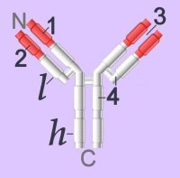
Immunoglobulins (left - click to enlarge) comprise two heavy (h) and two light-chain (l) protein subunits, each of which folds into domains (4 on heavy, 2 on light). These adhesion sites or domains contain one or more folds of 60 to 100 amino acids.

Depending upon the character of the heavy chain, immunoglobulins are divided into five classes – IgG, IgD, IgE, IgA, IgM – that are expressed in different tissues. The classes are further subdivided into isotypes, which have different properties in terms of complement fixation and binding to immunoglobulin (Ig) receptors.

Members of the immunoglobulin supergene family are found as:
● membrane-bound surface receptors of immune-system cells,
cellular adhesion molecules, or
● soluble antibodies (γ-globulins) synthesized by activated B cells.

Membrane-bound Igs have a transmembrane segment and a cytoplasmic C-terminal tail. The 2 β- chains are stabilized into sandwiched β sheets that are adherent by virtue of hydrophobic interactions between disulphide bonds. Igs assume a Y-shaped structure "topped" at the extracellular N-terminals by variable domains (red), with a variable domain at the tip of the heavy chain (1) and the light chain (2), between which lies an antigen binding site (3). The variable regions are coded by pluripotential DNA sequences that can generate thousands of polypeptide sequences capable of adhering to millions of different ligands. Binding is homophilic or heterophilic, including binding to different Igs and to integrins. Both light and heavy chains contain constant domains (white, 4).

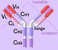
Right - click to enlarge - the heavy chains of IgA, IgD and IgG each have four domains, where those at the N-terminal are variable (VH) and the other three are constant (CH1-3). IgE and IgM have one variable and four constant domains (CH1-4) on the heavy chain. The variable domains are termed Fab, while the constant domains are termed Fc.

The light chains have two domains, one variable domain (VL) at the N-terminal, and one constant (CL) domain.

The antigen binding site lies between VH and VL (shaded lavendar). Most variability is found in three superficial-loop forming regions in the VH and VL domains, which are the complementarity determining regions or CDRs. CDR3 binds antigens and CDR1-2 bind MHCs. CDR3 shows more variation that do either CDR1 or 2.

The domains have related amino acid sequences that possess a common secondary and tertiary structure. This conserved structure is found frequently in proteins involved in cell-cell interactions and is particularly important in immunology. The constant (Fc) regions have complement fixing and Ig receptor binding activity. The hinge region, in IgG, IgA and IgD, is an important sequence of 10-60 amino acids between CH1 and CH2 that confers flexibility on the molecule.

animations Џ B cell selection Џ ELISA test +ve, -ve Џ IgG rotating x- y- axes Џ Rotating mouse IgG2a Molecule (y-axis) Џ somatic recombination of Ig gene Џ spinning IgG1 Kol Џ unfolding (small) IgG . unfolding (large) IgG .

Immunoglobulins attain their enormous variability by splicing components (VDJ recombination) coded in widely scattered sequences of DNA that are located in two different chromosomes. Antigen binding takes place at the heavy chain, which displays enormous variation by virtue of combining 1 of 400 possible variable gene segments with 1 out of 15 diversity segments and 1 out of 4 joining segments. This alternative splicing generates 24,000 possible combinations for the DNA encoding the heavy chain alone. The variable coding segments are assembled together with those for the constant-C segments of the heavy-chain molecule.

Tissue location:
IgA – mucus – gut, respiratory tract
IgD – antigen receptor on B cells
IgE – mast cells – releases histamines in response to allergens
IgG – primary immunity against invading pathogens
IgM – early B cell-mediated response to invading pathogens

Some antibody classes form multimeric structures – pentamers (IgM) and dimers or trimers (IgA). These two isotypes also associate with a small protein called the joining (J) chain required for stabilisation of the complexes.

The immunoglobulin superfamily is evolutionarily ancient, is widely expressed, and is constitutive or long-term up-regulated. Immunoglobulin antibodies are released by activated B cells of the immune system, on which they also act as surface marker proteins. Adherence of immunoglobilins to foreign substances or to cellular invaders may be sufficient to disarm the invader, or the attached antibodies function as attack signal to macrophages and natural killer cells. Adhesion molecules of the immunoglobulin supergene family, activate specific kinases through phosphorylation, resulting in activation of transcription factors, increased cytokine production, increased cell membrane protein expression, production of reactive oxygen species, and cell proliferation.

▲: adhesion molecules ~ adhesion molecules ф antigen : antigen binding site ф APCs ф B cells : C : CH1-4 : cellular adhesion molecules : complement fixation ф complement system : complementarity determining regions : constant domains : domains : evolution of immunoglobulins : Fab : Fc : heavy chain : hinge region ф humoral immunity : Ig supergene family ~ immunoglobulins : isotypes : kinase activationkinases : light chain : location of Ig classes : membrane-bound Igs : multimeric structures ф receptors ф signaling ф surface receptors ф T cells : tissue location ~ tyrosine kinases : V : VDJ recombination ф VDJ recombination : VH, VL : variable domains :▲

Tables  Fc receptors  Immune Cytokines  Immunoglobulins  Cell Adhesion Molecules  Cell signaling  Receptor Tyrosine Kinases (RTKs)  Receptor Signal Transduction  Second Messengers 

Top

tags

Labels: , , , , , , , , , ,

| 0 Guide-Glossary

antigen

An antigen is any molecule that stimulates an immune response. Most antigens are proteins or polysaccharides, though small molecules coupled to carrier proteins (haptens) can also be antigenic. The segment of an antigenic molecule to which its cognate antibody binds is termed an epitope or antigenic determinant.

allergen : allergic reactions : antigenic determinant : autoantigenic : autoimmune disorders : class I histocompatibility molecule (MHC I) : class II histocompatibility molecule (MHC II) : immunogen : endogenous : epitope : exogenous : lipid Ag : pathogen-associated molecular pattern (PAMP) : pattern-recognition receptor (PRR) : polysaccharide Ag : T-dependent : T-independent : tolerogen : Toll-like receptor (TLR) : tumor antigens : tumor-associated antigen (TAA) : tumor-specific antigen (TSA) ▼

Antigens are classified by immune activity as immunogens, tolerogens, or allergens according to whether the molecule in question activates the immune response, is tolerated by the immune system, or elicits an allergic response, respectively. Allergic reactions are exaggerated immune responses to molecules (allergens) that would otherwise not prove harmful. Antigens may also be classified according to their source as exogenous, endogenous, autoantigenic, or tumor antigens.

Exogenous antigens are foreign molecules that are ingested (endo-, phagocytosis) by antigen presenting cells on which the fragmented and extruded antigens are then carried on class II histocompatibility molecules (MHC II) for presentation to CD4+ Th cells. Pathogen-associated molecular patterns (PAMPs ) are small molecular sequences consistently found on pathogens that are recognized by Toll-like receptors (TLRs) and other pattern-recognition receptors (PRRs). Pattern recognition receptors (PRR) are a class of innate immune response-expressed protein receptors that respond to PAMPs.

Endogenous antigens are internally generated molecules that become presented on the cell surface in the complex with class I histocompatibility molecules (MHC I). Endogenous antigens may result from exogeneous viral or bacterial infections that have altered the host cell.

In autoimmune disorders, endogenous, self-molecules induce autimmune attack by CD8+ Tc/CTLs that have escaped negative selection in the thymus.

Tumor-specific antigens (TSAs) typically result from a tumor specific mutation and are targetted for non-self attack when displayed on class I histocompatibility molecules. Tumor-associated antigens (TAAs) are more common than TSAs, and are presented both by tumor cells and by normal cells. Tumor antigens may elicit targetting by CTLs before the tumor cells can successfully proliferate and metastasize. Unfortunately, tumors employ a variety of mechanisms to evade the immune system.

Protein antigens are T dependent in that they require T cell co-operation to induce antibody responses in B cells. Non-protein antigens, such as polysaccharides and lipids can elicit T-independent antibody responses. Such T-independent antigens are typically polymeric, so it is believed that they are able to cross-link BCR-surface-Ig sufficiently strongly to activate B cells without T cell costimulation. These T-independent polymeric antigens elicit IgM antibodies and do not demonstrate affinity maturation. However, a subclass of T cells are specialized to present lipid and glycolipid antigens – γδ T cells recognize foreign nonpeptide antigens presented by CD1 proteins, which are MHC-like-molecules specialized for the presentation of lipids.

ф activation ф anergy ф antibodies ф antigen presenting cells (APCs) ф autoimmunity ф basophils ¤ cancercell-cycle control ф class-switch recombination ф clonal selection ф dendritic cells o-o endogenous vs exogenous ф eosinophils ф granulocytes ~ growth factors ф immune cytokines ф immune response ф immune tolerance ф inflammatory response ф interferons ф isotype switching ф leukocytes ф lymphocytes ф lymphokines ф lymphoid system ф macrophages ф MHC ф monocytes ф pathogens ф pattern-recognition receptors ф phagocyte ф plasma cells ¤ proliferation ф receptorsregulation of gene expression ф secondary antibody diversification ф signaling ф surface receptors ф vaccines

Tables  Fc receptors  Immune Cytokines  Immunoglobulins  Cell Adhesion Molecules  Cell signaling  Receptor Tyrosine Kinases (RTKs)  Receptor Signal Transduction  Second Messengers 

Top

tags

Labels: , , , , , , , , , ,

| 0 Guide-Glossary

. . . since 10/06/06
Google