


killer T cells
Cytotoxic, CTLs, or killer T cells (CD8+) release lysis-inducing lymphotoxins.
▼ Apo_1 : apoptosis : C9 : CD8+ : CD95 : caspases : class I MHC : CIK cells : CTLs : cytotoxic T lymphocytes : epitope : Fas-FasL : granzymes : LAK cells : LAK anti-tumor Tx : membrane attack complex : MHC class I : natural killer cells : NK cells : NK receptors : perforin : perforin gene : perforin pore : serine protease : Tc : TIL cells : TNFR ▼
▼ Tc → CTL , some CTL → CIK
▼ natural killer cells (NK)
▼ laboratory: leukocyte + IL-2 → LAK
▼ anti-tumor Tx TIL, LAK
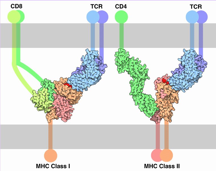
Tc cells express CD8 plus an antigen-specific T cell receptor (TCR) that ligates antigens presented by MHC class I, and are derived from a common lymphoid progenitor matured in the thymus. These Tc cells, once activated and matured into cytotoxic T lymphocytes (CTLs), recognize and destroy infected/altered self-cells.
Cytotoxic T lymphocytes (CTLs) and natural killer (NK) cells lyse target cells. CTLs recognize a specific epitope peptide presented by major histocompatibility complex (MHC) class I molecules on the target cells. NK cells recognize targets that express no class I MHC molecules I or low levels of MHC I molecules, enabling NK cells to target cells that have evaded CTLs.
Evidence suggests that both NK cells and CTLs exclusively employ perforin-dependent cytolysis. Perforin is a 534 aa glycoprotein with sequence homology to the membrane attack component of complement C9. As a cytolytic mediator, perforin integrates into the target cell membrane, forming polyprotein pores up to 20nm in diameter comprising 12—18 perforin monomers, which breach membrane integrity and permitting cytolytic cell death. The cell-type-specific expression of the perforin gene is determined by the promoter and upstream region, and there is evidence of transcriptional repression of the perforin gene in non-killer cells by two novel regulatory elements, one of which that is exclusively expressed by non-killer cells appears to be an Ets family member.[p]
Perforin pores in the plasma membrane of targetted cells permit entry of serine-protease granzymes, which cleave caspase precursors and trigger apoptosis. Perforin-dependent cytolysis plays an important role in immune response to bacterial and viral infections, in tumour surveillance, in immunopathology, and in autoimmunity.
A second cytolytic mechanism is mediated by the interaction of activated T cell surface FasL with TNFR family Fas receptors (TNFRSF6, Apo_1, CD95) expressed on the surface of the target cell. Fas and TNFR1 share a a conserved extracellular region and a cytoplasmic domain, crucial for the induction of apoptosis. Targets that express Fas can be lysed upon interaction with FasL on the surface of an effector cell (usually an activated T cell) or upon cross-linking by anti-Fas antibodies. A deficiency of functional Fas—FasL interactions results in lymphoproliferation, abnormal constitution of the peripheral T cell pool, and autoimmunity. [pdf]
Cytokine-induced killer (CIK) cells are a unique population of cytotoxic T lymphocytes (CTL) with the characteristic CD3+CD56+ phenotype. CIK cells also express CD94, part of the NK receptor comprising of CD94-NKG2 heterodimer. CIKs have demonstrated higher proliferative and cytolytic activities in comparison to the reported CD3-CD56+ lymphokine activated killer (LAK) cells that are essentially activated natural killer (NK) cells. CIK cells are non-MHC-restricted in target cell recognition and killing.
Lymphokine-activated killer cells (LAKs) are leukocytes stimulated in the laboratory to kill tumor cells. LAKs are generated by culturing spleen or peripheral blood leukocytes in presence of high concentrations of IL-2. LAKs are T-like cells that express CD3 or CD8. They lyse targets non-specifically, and with a target range hierarchy different than that of NK cells. LAK cells respond chemotactically and accumulate at sites of viral infection.
Natural killer (NK) cells are very important components of the innate immune system, are distinct from CTLs. NK, natural killer cells constitute a corps of circulating lymphocytes that are constitutively specialized to attack cancerous cells and virus infected cells. Preprogramming for target recognition, coupled with the absense of need for backup by a clone of identical cells, renders NK cells capable of rapid (innate) response to pathogens.
Individuals inherit multiple, polymorphic genes for NK receptors, so the assemblage of NK receptors differs between individuals. NK cells carry two forms of surface receptors:
● killer inhibitory receptors (KIR) belonging to the immunoglobulin superfamily. KIRs transmit an inhibitory signal when they encounter class I MHC molecules on a cell surface. (By contrast, T cells only recognize antigens that are presented by a MHC molecule.)
● activating receptors, which activate the NK cell upon binding to a target cell
-- ● killer lectin like receptor (KLR) CD94/NKG2 heterodimer, which employs MHC as ligand
-- ● natural cytotoxic receptors (NCR) which do not associate with MHC.
Lymphokine-activated killer (LAK) and tumor-infiltrating lymphocytes (TIL) have been employed therapeutically since the '70s mainly in end-stage patients with solid tumors. TIL are more specific and potent cytotoxic effectors than LAK, but their clinical use be considered potentially useful only in a few patients, chiefly those with solid tumors such as melanoma and glioblastoma.[r]
▲ Tc → CTL , some CTL → CIK
▲ natural killer cells (NK)
▲ laboratory: leukocyte + IL-2 → LAK
▲ anti-tumor TIL, LAK
▲ ф antibodies ф antigen : Apo_1 : apoptosis סּ apoptosis ф B cells : C9 ф C9: CD8+ : CD95 ф CD : caspases : class I MHC ф complement system ф classes of MHC : CTLs ф cytolysis : cytotoxic T lymphocytes : epitope : Fas-FasL ф granulocytes : granzymes : membrane attack complex ф membrane attack complex ф MHC : MHC class I : natural killer cells ф : NK cells ф NK cells : perforin : perforin gene : perforin pore ф C9 pore ф receptors : serine protease ф serine proteases ф surface receptors : Tc : TNFR ▲
▲ Top ▲
Tables Complement Receptors Fc receptors Immune Cytokines Immunoglobulins Interferons Scavenger Receptors Toll-like Receptors Cell Adhesion Molecules Cell signaling Receptor Tyrosine Kinases (RTKs) Receptor Signal Transduction Second Messengers

▼ Apo_1 : apoptosis : C9 : CD8+ : CD95 : caspases : class I MHC : CIK cells : CTLs : cytotoxic T lymphocytes : epitope : Fas-FasL : granzymes : LAK cells : LAK anti-tumor Tx : membrane attack complex : MHC class I : natural killer cells : NK cells : NK receptors : perforin : perforin gene : perforin pore : serine protease : Tc : TIL cells : TNFR ▼
▼ Tc → CTL , some CTL → CIK
▼ natural killer cells (NK)
▼ laboratory: leukocyte + IL-2 → LAK
▼ anti-tumor Tx TIL, LAK
Tc cells express CD8 plus an antigen-specific T cell receptor (TCR) that ligates antigens presented by MHC class I, and are derived from a common lymphoid progenitor matured in the thymus. These Tc cells, once activated and matured into cytotoxic T lymphocytes (CTLs), recognize and destroy infected/altered self-cells.
Cytotoxic T lymphocytes (CTLs) and natural killer (NK) cells lyse target cells. CTLs recognize a specific epitope peptide presented by major histocompatibility complex (MHC) class I molecules on the target cells. NK cells recognize targets that express no class I MHC molecules I or low levels of MHC I molecules, enabling NK cells to target cells that have evaded CTLs.
Evidence suggests that both NK cells and CTLs exclusively employ perforin-dependent cytolysis. Perforin is a 534 aa glycoprotein with sequence homology to the membrane attack component of complement C9. As a cytolytic mediator, perforin integrates into the target cell membrane, forming polyprotein pores up to 20nm in diameter comprising 12—18 perforin monomers, which breach membrane integrity and permitting cytolytic cell death. The cell-type-specific expression of the perforin gene is determined by the promoter and upstream region, and there is evidence of transcriptional repression of the perforin gene in non-killer cells by two novel regulatory elements, one of which that is exclusively expressed by non-killer cells appears to be an Ets family member.[p]
Perforin pores in the plasma membrane of targetted cells permit entry of serine-protease granzymes, which cleave caspase precursors and trigger apoptosis. Perforin-dependent cytolysis plays an important role in immune response to bacterial and viral infections, in tumour surveillance, in immunopathology, and in autoimmunity.
A second cytolytic mechanism is mediated by the interaction of activated T cell surface FasL with TNFR family Fas receptors (TNFRSF6, Apo_1, CD95) expressed on the surface of the target cell. Fas and TNFR1 share a a conserved extracellular region and a cytoplasmic domain, crucial for the induction of apoptosis. Targets that express Fas can be lysed upon interaction with FasL on the surface of an effector cell (usually an activated T cell) or upon cross-linking by anti-Fas antibodies. A deficiency of functional Fas—FasL interactions results in lymphoproliferation, abnormal constitution of the peripheral T cell pool, and autoimmunity. [pdf]
Cytokine-induced killer (CIK) cells are a unique population of cytotoxic T lymphocytes (CTL) with the characteristic CD3+CD56+ phenotype. CIK cells also express CD94, part of the NK receptor comprising of CD94-NKG2 heterodimer. CIKs have demonstrated higher proliferative and cytolytic activities in comparison to the reported CD3-CD56+ lymphokine activated killer (LAK) cells that are essentially activated natural killer (NK) cells. CIK cells are non-MHC-restricted in target cell recognition and killing.
Lymphokine-activated killer cells (LAKs) are leukocytes stimulated in the laboratory to kill tumor cells. LAKs are generated by culturing spleen or peripheral blood leukocytes in presence of high concentrations of IL-2. LAKs are T-like cells that express CD3 or CD8. They lyse targets non-specifically, and with a target range hierarchy different than that of NK cells. LAK cells respond chemotactically and accumulate at sites of viral infection.
Natural killer (NK) cells are very important components of the innate immune system, are distinct from CTLs. NK, natural killer cells constitute a corps of circulating lymphocytes that are constitutively specialized to attack cancerous cells and virus infected cells. Preprogramming for target recognition, coupled with the absense of need for backup by a clone of identical cells, renders NK cells capable of rapid (innate) response to pathogens.
Individuals inherit multiple, polymorphic genes for NK receptors, so the assemblage of NK receptors differs between individuals. NK cells carry two forms of surface receptors:
● killer inhibitory receptors (KIR) belonging to the immunoglobulin superfamily. KIRs transmit an inhibitory signal when they encounter class I MHC molecules on a cell surface. (By contrast, T cells only recognize antigens that are presented by a MHC molecule.)
● activating receptors, which activate the NK cell upon binding to a target cell
-- ● killer lectin like receptor (KLR) CD94/NKG2 heterodimer, which employs MHC as ligand
-- ● natural cytotoxic receptors (NCR) which do not associate with MHC.
Lymphokine-activated killer (LAK) and tumor-infiltrating lymphocytes (TIL) have been employed therapeutically since the '70s mainly in end-stage patients with solid tumors. TIL are more specific and potent cytotoxic effectors than LAK, but their clinical use be considered potentially useful only in a few patients, chiefly those with solid tumors such as melanoma and glioblastoma.[r]
▲ Tc → CTL , some CTL → CIK
▲ natural killer cells (NK)
▲ laboratory: leukocyte + IL-2 → LAK
▲ anti-tumor TIL, LAK
▲ ф antibodies ф antigen : Apo_1 : apoptosis סּ apoptosis ф B cells : C9 ф C9: CD8+ : CD95 ф CD : caspases : class I MHC ф complement system ф classes of MHC : CTLs ф cytolysis : cytotoxic T lymphocytes : epitope : Fas-FasL ф granulocytes : granzymes : membrane attack complex ф membrane attack complex ф MHC : MHC class I : natural killer cells ф : NK cells ф NK cells : perforin : perforin gene : perforin pore ф C9 pore ф receptors : serine protease ф serine proteases ф surface receptors : Tc : TNFR ▲
▲ Top ▲
Tables Complement Receptors Fc receptors Immune Cytokines Immunoglobulins Interferons Scavenger Receptors Toll-like Receptors Cell Adhesion Molecules Cell signaling Receptor Tyrosine Kinases (RTKs) Receptor Signal Transduction Second Messengers
Labels: CIK, CTL, cytotoxic, epitope protein, granzymes, killer T cells, LAK, MHC class I protein, natural killer, perforin, T lymphocytes
| 0 Guide-Glossary